Page 338 - Cardiac Nursing
P. 338

Pa
                                           g
                                        M
                                          Pa
                                            e 3
                                            e 3
                                           g
                                           g
                                        M
                                   1
                                   1
                                /09
                                /09
                                     0 P
                                     0 P
                                   0:3
                                   0:3
                                                   a
                                                   a
                                                  ara
                                                  ara
                                                      c.
                                                      c.
                                                    In
                                                    In
                                                  t
                                                A
                                                A
                                              14
                                              14
                                                 p
                                                  t
                                                 p
                                                 p
                              /29
         LWB
                      0-3
            K34
                        32.
               0-c
                      0-3
         LWBK340-c15_
         LWB K34 0-c 15_ p pp300-332.qxd  6/29/09  10:30 PM  Page 314 Aptara Inc.
                   p
                    30
                    30
                             6
                           xd
                 15_
                              /29
                             6
                          q
                        32.
                          q
                           xd
                          q
                  314    P A R T  III / Assessment of Heart Disease
                                                         ead I
                                                         Le
                                 Anterior fascicle                                                       I I I
                      Posterior
                       fascicle
                                  Lead III
                                                                                  III III III I
                                                                      B
                   A
                                                 I I I
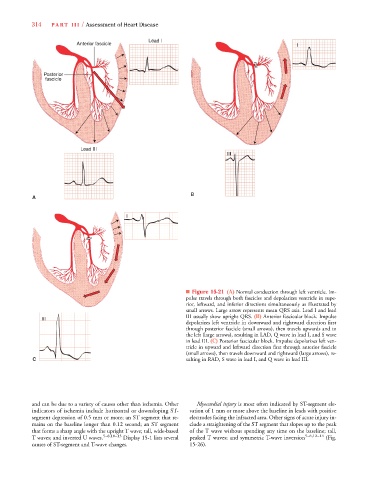
                                                                     ■ Figure 15-21 (A) Normal conduction through left ventricle. Im-
                                                                     pulse travels through both fascicles and depolarizes ventricle in supe-
                                                                     rior, leftward, and inferior directions simultaneously as illustrated by
                                                                     small arrows. Large arrow represents mean QRS axis. Lead I and lead
                                                                     III usually show upright QRS. (B) Anterior fascicular block. Impulse
                      III III III I I
                                                                     depolarizes left ventricle in downward and rightward direction first
                                                                     through posterior fascicle (small arrows), then travels upwards and to
                                                                     the left (large arrows), resulting in LAD, Q wave in lead I, and S wave
                                                                     in lead III. (C) Posterior fascicular block. Impulse depolarizes left ven-
                                                                     tricle in upward and leftward direction first through anterior fascicle
                                                                     (small arrows), then travels downward and rightward (large arrows), re-
                   C                                                 sulting in RAD, S wave in lead I, and Q wave in lead III.


                  and can be due to a variety of causes other than ischemia. Other  Myocardial injury is most often indicated by ST-segment ele-
                  indicators of ischemia include horizontal or downsloping ST-  vation of 1 mm or more above the baseline in leads with positive
                  segment depression of 0.5 mm or more; an ST segment that re-  electrodes facing the infracted area. Other signs of acute injury in-
                  mains on the baseline longer than 0.12 second; an ST segment  clude a straightening of the ST segment that slopes up to the peak
                  that forms a sharp angle with the upright T wave; tall, wide-based  of the T wave without spending any time on the baseline; tall,
                  T waves; and inverted U waves. 5–8,10–13  Display 15-1 lists several  peaked T waves; and symmetric T-wave inversion 5–8,12–14  (Fig.
                  causes of ST-segment and T-wave changes.            15-26).
   333   334   335   336   337   338   339   340   341   342   343