
Page 339 - Cardiac Nursing
P. 339
0 P
M
0 P
0:3
0:3
g
g
Pa
M
Pa
6
/29
6
xd
xd
1
1
/09
/29
/09
p
p
t
ara
t
15
15
A
p
A
c.
c.
e 3
g
e 3
a
ara
a
In
In
q
0-3
0-3
K34
LWBK340-c15_
p
p
30
30
32.
32.
q
q
15_
0-c
LWB K34 0-c 15_ pp300-332.qxd 6/29/09 10:30 PM Page 315 Aptara Inc.
LWB
C HAPTER 1 5 / Electrocardiography 315
R
R R R R R R R R
V V V V V
VR
VR
VR
aV
aV
I I I I aV R V V V V V V V V V V V V 1 1 1 1 1 1 1 V V V V V V V V V V V V 4 4 4 4 4 4 4
aV
a a a a a a a a a a
V
V
VRR
a a a a a a a a
aV
aV
I I I I I I I I aV L L L L L L L L L V V V V V V V V V V V V V V V 2 2 2 2 2 2 2 V V V V V V V V V V V V V V V 5 5 5 5 5 5 5
V
VLL
VL
VL
V V V V V
VL
V
V
F
VF
VFF
VF
VF
II II II II I I I I aV F F F F V V V V V V V V V V V V V V V
V
V
V
V
V V V V V V V V
aV
a a a a a
aV
V V V V V V V V V V V V V V V 3 3 3 3 3 3 3 3 3 6 6 6 6 6 6 6
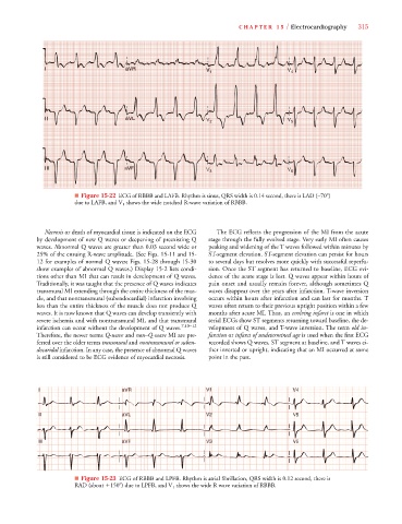
■ Figure 15-22 ECG of RBBB and LAFB. Rhythm is sinus, QRS width is 0.14 second, there is LAD (–70 )
due to LAFB, and V 1 shows the wide notched R-wave variation of RBBB.
Necrosis or death of myocardial tissue is indicated on the ECG The ECG reflects the progression of the MI from the acute
by development of new Q waves or deepening of preexisting Q stage through the fully evolved stage. Very early MI often causes
waves. Abnormal Q waves are greater than 0.03 second wide or peaking and widening of the T waves followed within minutes by
25% of the ensuing R-wave amplitude. (See Figs. 15-11 and 15- ST-segment elevation. ST-segment elevation can persist for hours
12 for examples of normal Q waves; Figs. 15-28 through 15-30 to several days but resolves more quickly with successful reperfu-
show examples of abnormal Q waves.) Display 15-2 lists condi- sion. Once the ST segment has returned to baseline, ECG evi-
tions other than MI that can result in development of Q waves. dence of the acute stage is lost. Q waves appear within hours of
Traditionally, it was taught that the presence of Q waves indicates pain onset and usually remain forever, although sometimes Q
transmural MI extending through the entire thickness of the mus- waves disappear over the years after infarction. T-wave inversion
cle, and that nontransmural (subendocardial) infarction involving occurs within hours after infarction and can last for months. T
less than the entire thickness of the muscle does not produce Q waves often return to their previous upright position within a few
waves. It is now known that Q waves can develop transiently with months after acute MI. Thus, an evolving infarct is one in which
severe ischemia and with nontransmural MI, and that transmural serial ECGs show ST segments returning toward baseline, the de-
infarction can occur without the development of Q waves. 7,10–12 velopment of Q waves, and T-wave inversion. The term old in-
Therefore, the newer terms Q-wave and non–Q-wave MI are pre- farction or infarct of undetermined age is used when the first ECG
Q
Q
ferred over the older terms transmural and nontransmural or suben- recorded shows Q waves, ST segment at baseline, and T waves ei-
docaridal infarction. In any case, the presence of abnormal Q waves ther inverted or upright, indicating that an MI occurred at some
is still considered to be ECG evidence of myocardial necrosis. point in the past.
V11
V V V V V V V V V V V V V V V
V1
V1
VR
VR
V44
V4
VR
V
V V V V V V V V
VR
V
V V V V4
R
R R R R R R R R
I I I I I a a a a a a a aV R V1 1 1 1 1 V4 4
R
V2
V22
V V V V V V V V
a
II II II II I a a a a a a a a aV L L L L L V V V V V V V V V V V V V V V 2 2 2 2 V V V5 5 5
V5
V55
VL
V V2
V5
VL
V
VL
VL
V
V6
V6
V3
V V V V V3
V3
V V V V6
V V V V6
V33
V V V V6
V3
V66
VF
a aV
VF
V
aVVF
III I I III II III II III II III III III II I I a aV F V3 3 3 3 3 V V V V6 6 6 6 6 6 6 6
a a aVF
aVF
VF
F
F
F
F
VF
V
V
V
■ Figure 15-23 ECG of RBBB and LPFB. Rhythm is atrial fibrillation, QRS width is 0.12 second, there is
RAD (about
150 ) due to LPFB, and V 1 shows the wide R wave variation of RBBB.