Page 340 - Cardiac Nursing
P. 340

1
                                   1
                                /09
                              /29
                                /09
                                   0:3
                                        M
                                        M
                                     0 P
                                   0:3
                                     0 P
                              /29
                        32.
                          q
                        32.
                      0-3
                      0-3
                          q
                             6
                             6
                           xd
                          q
                           xd
                                          Pa
                                                  ara
                                                  ara
                                                  t
                                                 p
                                                  t
                                                   a
                                                      c.
                                                      c.
                                                    In
                                                   a
                                                    In
                                                 p
                                           g
                                            e 3
                                           g
                                          Pa
                                           g
                                            e 3
                                                A
                                                 p
                                                A
                                              16
                                              16
                    30
         LWB K34 0-c 15_ pp300-332.qxd  6/29/09  10:30 PM  Page 316 Aptara Inc.
                   p
         LWB
         LWBK340-c15_
               0-c
                   p
                 15_
                    30
            K34
                  316    P A R T  III / Assessment of Heart Disease
                   Reciprocal                          Indicative                    Causes of St-Segment and T-Wave
                                                                                                 g
                                                                                                 g
                   Changes                             Changes         DISPLAY 15-1        2,3,5–7,10–12,14–16
                                                                                     Changes
                                                   Ischemia            Aberrant conduction   Hypothermia
                                                                       Apical ballooning     Intracranial hemorrhage
                                                                         syndrome (Takotsubo)  Myocardial metastases
                                                   Injury              Amyloidosis           Myocarditis
                                                                       Bundle-branch block   Paced rhythm
                                                                       Cardiomyopathy        Pancreatitis or acute abdomen
                                                   Necrosis
                                                                       Cocaine vasospasm     Pericarditis
                                                                       Drugs                 Physical training
                    Non-Q-wave                     Healthy             Early repolarization  Prinzmetal’s angina
                    Infarction                     Tissue              Hemiblock             Pulmonary embolism
                                                                       Hypercalcemia         Tachycardia
                                                                       Hyperkalemia          Ventricular aneurysm
                                                                       Hyperventilation      Ventricular hypertrophy
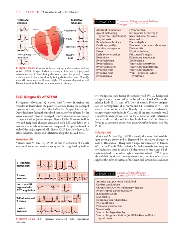
                  ■ Figure 15-24 Zones of ischemia, injury, and infarction with as-
                  sociated ECG changes. Indicative changes of ischemia, injury, and  Hypocalcemia  Ventricular rhythms
                                                                       Hypoglycemia
                                                                                             Wolff–Parkinson–White
                  necrosis are seen in leads facing the injured area. Reciprocal changes  Hypokalemia  syndrome
                  are often seen in leads not directly facing the involved area. Non–Q-
                  wave MI causes reduced R wave height, ST segment depression, and
                  T-wave inversion. Ischemia can also present this way.
                  ECG Diagnosis of STEMI                              tive changes in leads facing the anterior wall (V 1–4 ). Reciprocal
                                                                      changes are often recorded in the lateral leads I and aVL and the
                  ST-segment elevation, Q waves, and  T-wave inversion are  inferior leads II, III, and aVF. Loss of normal R-wave progres-
                  recorded in leads where the positive electrode facing the damaged  sion or development of Q waves and ST elevation in V 1–4 are
                  myocardium and are called the indicative changes of infarction.  seen in anterior infarction. If only the septum is infarcted,
                  Other leads not facing the involved tissue are often affected by the  changes occur only in leads V 1–2 , but, if the entire anterior wall
                  loss of electrical forces in damaged tissue and record mirror-image  is involved, changes are seen in V 1–4 . Anterior wall infarction
                  changes called reciprocal changes. Figure 15-24 illustrates indica-  that extends laterally and involves leads I and aVL is often re-
                  tive and reciprocal changes associated with MI, and Table 15-1  ferred to as extensive anterior or anterolateral infarction (see Fig.
                  lists leads in which indicative and reciprocal changes are found in  15-31).
                  each of the major types of MI. Figure 15-27 illustrates how to lo-
                  calize ischemia, injury, and infarction using the 12-lead ECG.  Inferior MI
                                                                      Inferior wall MI (see Fig. 15-29) is usually due to occlusion of the
                  Anterior MI                                         right coronary artery and is diagnosed by indicative changes in
                  Anterior wall MI (see Fig. 15-28) is due to occlusion of the left  leads II, III, and aVF. Reciprocal changes are often seen in leads I,
                  anterior descending coronary artery and is recognized by indica-  aVL, or the V leads. When inferior MI is due to right coronary ar-
                                                                      tery occlusion, there is usually ST depression in lead I and ST el-
                                                                      evation in lead III, which is higher than that in lead II. 7,10  In peo-
                                                                      ple with left dominant coronary circulation, the circumflex artery
                                                                      supplies the inferior surface of the heart and circumflex occlusion
                   ST segment
                   depression
                                                                       DISPLAY 15-2  Causes of Noninfarction
                   T wave                                                            Q Waves 2,3,5–7,10–12,14–16
                   inversion
                                                                       Anterior and posterior hemiblock
                   Horizontal ST                                       Cardiac amyloidosis
                   segment with                                        Chronic obstructive pulmonary disease
                   ST-T angulation                                     Hypertrophic cardiomyopathy
                                                                       Incomplete LBBB
                                                                       Myocarditis
                   Tall wide-based
                   T waves                                             Neuromuscular disorders
                                                                       Pneumothorax
                                                                       Pulmonary embolism
                   U wave                                              Sarcoidosis
                   inversion
                                                                       Ventricular hypertrophy
                                                                       Ventricular preexcitation (Wolff–Parkinson–White
                  ■ Figure 15-25 ECG  patterns associated with myocardial   syndrome)
                  ischemia.
   335   336   337   338   339   340   341   342   343   344   345