
Page 341 - Cardiac Nursing
P. 341
LWBK340-c15_p300-332.qxd 6/29/09 10:30 PM Page 317 Aptara Inc.
C HAPTER 1 5 / Electrocardiography 317
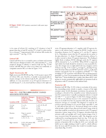
ST elevation 1 mm or
more in two
contiguous leads
ST pulled up to peak
of T wave with no J
■ Figure 15-26 ECG patterns associated with acute myo- point
cardial injury.
Tall, peaked T waves
Symmetrical T wave
inversion
is the cause of inferior MI, resulting in ST elevation in lead II tricle. ST-segment elevation in V 1 together with ST-segment ele-
greater than that in lead III, and the ST in lead I is either isoelec- vation in the inferior leads is suspect for RVMI. Another clue is
tric or elevated. 10 Approximately 30% of inferior MIs involve the discordance between the ST segment in V 1 and the ST segment
V
right ventricle 7,17 (see Fig. 15-32). in V 2 . Discordance means that the ST segments do not point in
V
the same direction—V 1 shows ST-segment elevation, whereas V 2
Lateral MI is either normal or shows ST-segment depression. This finding is
Lateral wall MI is due to circumflex artery occlusion and presents highly likely to indicate RVMI, although rarely the ST segment
V
with indicative changes in leads I, aVL, and sometimes V 5–6 , with will be elevated in V 1 –V 4 in RVMI. When RVMI is suspected,
reciprocal changes in inferior or anterior leads (see Fig. 15-30). right-sided chest leads should be obtained as soon as possible be-
Lateral wall MI does not often occur alone but commonly ac- cause the changes seen in right-sided leads may disappear within
V
companies anterior MI, as it does in Figure 15-31. 24 hours (see Fig. 15-9). Leads V 4R through V 6R develop ST-seg-
ment elevation when acute RVMI is present. Lead V 4R is the most
V
sensitive and specific lead for recognition of RVMI. 7,10,17 The
Right Ventricular MI recording of V 4 R in patients with inferior MI and hemodynamic
V
Right ventricular MI (RVMI; see Fig. 15-32) occurs in up to 45% instability is a class I recommendation from the 2004 ACC/AHA
of inferior MIs, and, therefore, it usually is associated with in- Task Force. 18 Some facilities have a policy that directs ECG tech-
dicative changes in the inferior leads II, III, and aVF. 7,10,11,17 In nicians to obtain automatically right-sided and posterior leads in
all patients with ST elevation in the inferior leads.
addition, it is not uncommon to see ST-segment elevation in V 1
as well, because V 1 is the chest lead that is closest to the right ven-
Posterior MI
Posterior wall MI (Fig. 15-33) is due to occlusion of the poste-
rior descending artery, which is usually a branch of the right
coronary artery. In left dominant circulations the posterior de-
Table 15-1 ■ ELECTROCARDIOGRAPHIC CHANGES scending is a branch of the circumflex artery. Isolated posterior
ASSOCIATED WITH STEMI wall MI is uncommon; it usually accompanies inferior or lateral
wall MI, 2,6,7,11 as seen in Figure 15-33B. ECG changes of poste-
Indicative Changes: Reciprocal Changes:
ST Elevation, ST Depression, Tall R rior MI are less obvious because in the standard 12-lead ECG
Q Waves, T Wave Waves, Upright there are no leads those face the posterior wall, and, therefore, no
Location of MI Inversion T Waves
indicative changes are recorded. The diagnosis is made by ob-
serving reciprocal changes in the anterior leads, especially V 1 and
Anterior V 1 to V 4 I, aVL, II, III, aVF
V
V 2 , but often all the way to V 4 . Reciprocal changes seen in these
Septal V 1 , V 2 I, aVL V V
V
Inferior II, III, aVF I, aVL, leads include a taller R wave than normal (mirror image of the Q
If seen in V 1 to V 3 suspect wave that would be recorded over the posterior wall), ST-segment
posterior MI
V
Posterior None in standard V 1 to V 4 depression (mirror image of the ST-segment elevation from the
12 leads posterior wall), and upright, tall T waves (mirror image of the T-
V V wave inversion from the posterior wall). The diagnosis can be
Posterior leads V 7 –V 9
Lateral I, aVL, V 5 , V 6 II, III, aVF confirmed by recording posterior leads (see Fig. 15-9) and ob-
If seen in V 1 , V 3 suspect serving ST elevation and Q waves. Another way to verify the
posterior MI
Right ventricle II, III, aVF (inferior MI) presence of posterior MI is to flip the ECG over vertically and
V
Right chest leads hold it up to a light, looking at leads V 1 and V 2 , which will now
V 4 R–V 6 R show Q waves and ST elevation that would be recorded in pos-
V
terior leads (Fig. 15-33C).