
Page 343 - Cardiac Nursing
P. 343
1
/09
/09
1
0 P
0:3
0:3
/29
p
p
xd
p
/29
6
6
A
e 3
e 3
A
19
19
c.
g
M
M
0 P
Pa
g
g
Pa
30
30
In
p
a
a
0-3
0-3
In
K34
0-c
LWBK340-c15_ p pp300-332.qxd 6/29/09 10:30 PM Page 319 Aptara Inc.
K34
15_
c.
0-c
15_
ara
32.
q
32.
q
t
ara
xd
q
t
C HAPTER 1 5 / Electrocardiography 319
VR
VR
a a a a a a a aV R
V V V V V
VR
VRR
R R R R R R R R
R
V4
V V V V V V V
V4
V
I I I I V V V V V V V V V V V V V V V 1 V4 4 4 4 4 4 4 4 4
V1
V1
V1
V1
V2
V V V V V V V V V V V V V V V
V2 2 2 2 2 2 2
V2
V2
V5
V5
V
V5
I I I I I I I I V V V V V V V V V 5 5 5 5 5 5 5 5 5 5
V5
VL
a a a a a a a aV L L L L L L L L L
VLL
VL
V V V V V
VL
V3
V3
V V V V V V V V V V V V V V V
V3
V6
V V V V V V V
II II II I I I a a a a a aV F F F F F F F F V3 3 3 3 3 3 3 V6 6 6 6 6 6 6 6 6 6 6 6
V
V6
a a
F
VF
VF
V V V V V
VFF
VF
C
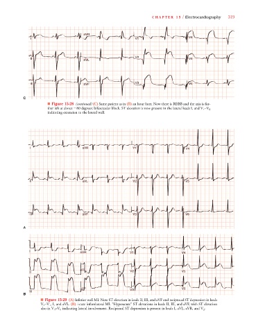
■ Figure 15-28 (continued) (C) Same patient as in (B) an hour later. Now there is RBBB and the axis is fur-
V
ther left at about 80 degrees: bifascicular block. ST elevation is now present in the lateral leads I, and V 4 –V 6
indicating extension to the lateral wall.
V11
V1
aV
a a a a a a a a
aV
V
V
I I I I aV R V1 1 1 1 1 V V V V V V V V V V V V V 4 4 4 4 4 4 4 4 4
V V V VR
V
V
V
V
R R R R R R R R R R R R
V1
V V V V V V V V V V
V4
V4
V
V
V4
V4
II II II II aV L L L L L L L L V V V V V V V V V V 2 2 2 2 2 2 2 2 2 V V V V V V V V V V V V V V 5 5 5 5 5 5 5 5
V V V VL
V
V
V
a a a a a a a a
aV
V
aV
V2
V22
V2
V2
V
V
V3
V V V V V V V V V V
V33
V3
V
V
aV
aV
V
V
V
V
V V V VF
a a a a a a a a
I I I I III III III III I I I I I I I I I I aV F F F F F F F F F F F F F V3 3 3 3 3 3 3 3 3 3 V V V V V V V V V V V V V V 6 6 6 6 6 6 6 6
A
I I I I I aV R R R R R R R R R R R R V1 1 1 1 1 1 V44 4 4 4 4 4
a a a a a a a a
aV
V
V V V VR
aVV
V
R
V4
V4
V4
V V V V V V V V V V
V4
V1
V1
V1
V V V V V V V V V V V
V
V
aVV
a a a a a a a a a a a a a a aV L L L L L L L L L L L L L L L L L
V V V VL
aV
V5
V5
V2
V5
V V V V V V V V V V
V55
V2
V V V V V V V V V V V
I I I I II II II II II V V2 2 2 2 2 2 2 2 2 V5 5 5 5 5 5
V
V
aV F F F F F F F F F F F F F F V V V V V V V V V 3 3 3 3 3 3 3 3 V6 6 6 6 6 6
V V V VF
F
F
V
F
aVV
V
V6
a a a a a a a a
V
V66
aV
V V V V V V V V V V V
V6
V3
V3
V3
I I I I II II II II II II II II I I I
B
■ Figure 15-29 (A) Inferior wall MI. Note ST elevation in leads II, III, and aVF and reciprocal ST depression in leads
V V
V 2 –V 4 , I, and aVL. (B) Acute inferolateral MI. “Hyperacute” ST elevations in leads II, III, and aVF, with ST elevation
V
V
also in V 4 –V 6 indicating lateral involvement. Reciprocal ST depression is present in leads I, aVL, aVR, and V 2 .