
Page 344 - Cardiac Nursing
P. 344
Pa
Pa
M
M
g
e 3
g
g
0 P
/09
1
/29
/09
0:3
0 P
1
0:3
e 3
t
p
ara
t
A
A
p
p
ara
c.
c.
20
20
a
a
In
In
0-c
15_
15_
q
q
0-c
LWBK340-c15_ p p pp300-332.qxd 6/29/09 10:30 PM Page 320 Aptara Inc.
30
0-3
0-3
30
32.
32.
/29
LWB
K34
LWB
K34
xd
xd
6
6
q
V11
VR
R R R R R R R R
a a
VR
R
V V V V V V V V V V
V1
V4
V4
V4
VR
I I I I I I I I I a a a a a aV V V V V V V VRR V1 1 1 1 1 V V V V V V V V V V V 4 4 4 4 4 4
V4
V1
VL
V5
V2
VLL
V22
V V V V V V V V
V5
II II III I I I I a a a a a a a aV V V V V V VL L L L L L L L V2 2 2 2 2 V V V V V V V V V 5 5 5 5 5
V5
V
VF
V V V V
V3
V V V V V V V V
V6
V V V V V V V V V
V6
III III III I I I I II a a a a a aV VFF V3 3 3 3 3 V6 6 6 6 6 6
V33
F
VF
a a
F F F F F F
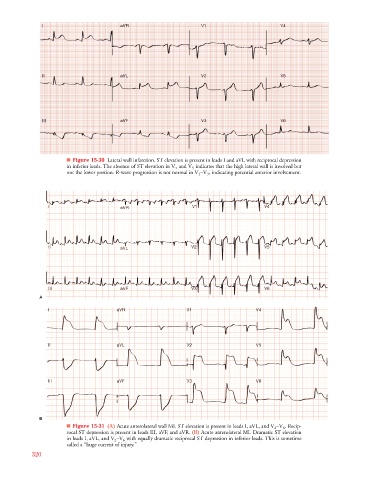
■ Figure 15-30 Lateral wall infarction. ST elevation is present in leads I and aVL with reciprocal depression
in inferior leads. The absence of ST elevation in V 4 and V 5 indicates that the high lateral wall is involved but
V
not the lower portion. R-wave progression is not normal in V 1 –V 3 , indicating potential anterior involvement.
V4
V V V V V V
V4
I I I I aV V V V V V V VR V V V V V V V V V V V V V V 1 1 1 1 1 V4 4 4 4 4 4 4 4 4 4 4
V4
aVV
a a a a a a a a
aV
R R R R R R R R
R
R
VRR
VR
VR
V5
V5
V2
V5
V V V V V V
V2
II II II III aV V V V V V V VL L L L L L V2 2 2 2 2 2 2 V5 5 5 5 5 5 5 5 5 5 5
V2
V V V V V V V V V V V V V V V
a a a a a a a a
aV
aVV
VLL
VL
VL
V6
V6
V3
V3
V V V V V V V V V V V V
I I I I II II II II I aV V V V V V V VF V3 3 3 3 3 3 3 3 V6 6 6 6 6 6 6 6 6 6 6
VF
VF
VFF
aV
a a a a a a a a
aVV
V6
V V V V V V
F F F F
F
A
V
V V V V V V V V
VR
V
V
I I I I I I I I I a a a a aV VR V V V V V V V V V V V V V V 1 1 1 1 1 V V V V V V V V V V4 4 4 4 4 4 4 4 4 4 4 4 4 4
VR
VRR
R
R R R R R R R R R
VL
V
I I I I II II II II I I I II I a a a a aV V V V V V V V V VL L L L L L V V V V V V V V V V V V V V 2 2 2 2 2 V V V V V V V V V V5 5 5 5 5 5 5 5 5 5 5 5 5 5
V
VLL
VL
V
F F F F F
II II II III II II II II I I I II I a a a a aV V V V V V V V V V V VFF V V V V V V V V V V V V V V 3 3 3 3 3 V V V V V V V V V V6 6 6 6 6 6 6 6 6 6 6 6 6 6
VF
VF
V
VF
B
■ Figure 15-31 (A) Acute anterolateral wall MI. ST elevation is present in leads I, aVL, and V 2 –V 6 . Recip-
V
rocal ST depression is present in leads III, aVF, and aVR. (B) Acute anterolateral MI. Dramatic ST elevation
V
in leads I, aVL, and V 2 –V 6 with equally dramatic reciprocal ST depression in inferior leads. This is sometime
called a “huge current of injury.”
320