
Page 348 - Cardiac Nursing
P. 348
1
0:3
1
/09
/09
0:3
M
Pa
M
0 P
0 P
q
q
q
32.
32.
xd
/29
/29
6
xd
6
Pa
ara
ara
t
p
t
a
c.
c.
In
a
In
e 3
e 3
g
g
g
24
p
p
A
24
A
p
30
K34
15_
0-c
30
0-3
0-3
LWB K34 0-c 15_ p pp300-332.qxd 6/29/09 10:30 PM Page 324 Aptara Inc.
LWBK340-c15_
LWB
324 P A R T III / Assessment of Heart Disease
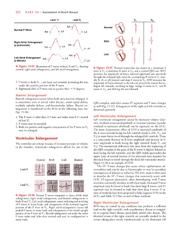
V
Lead 11 Lead VV 1 V 1 V 6
RA Normal
Normal P Wave
RA LA LA
Right Atrial Enlargement RVH
(p pulmonale) RA
RA LA
LA
RA LVH
Left Atrial Enlargement RA LA
(p Mitrale) LA
■ Figure 15-35 Illustration of P waves in leads II and V 1 , showing ■ Figure 15-37 Normal ventricular size results in a dominant S
normal, right atrial enlargement, and left atrial enlargement.
wave in V 1 , a dominant R wave in V 6 , and a normal QRS axis. RVH
increases the amplitude of forces directed rightward and anteriorly
through the enlarged right ventricle, causing large R waves in V 1 (usu-
ally R, rS, or qR pattern) and deep S waves in V 6 . LVH increases the
2. P waves in leads V 1–3 are sharp and pointed, increasing the area amplitude of forces directed to the left and posteriorly toward the en-
under the positive portion of the P wave. larged left ventricle, resulting in large voltage S waves in V 1 and R
3. Rightward shift of P wave axis to greater than
75 degrees. waves in V 6 , and shifting the axis leftward.
Biatrial Enlargement
Biatrial enlargement occurs when both atria become enlarged. It
is sometimes seen in mitral valve disease, atrial septal defect, QRS complex, and often causes ST segment and T wave changes
multiple valvular defects, and biventricular failure. Biatrial en- as well (Fig. 15-37). Enlargement of the right and left ventricles is
largement is manifested on the ECG in the following ways (see discussed separately.
Fig. 15-36):
1. The P wave is taller than 2.5 mm and wider than 0.11 second Left Ventricular Enlargement
in lead II. Left ventricular enlargement caused by increased volume (dias-
2. P waves may be notched. tolic overload or increased preload) or increased pressure (systolic
overload or increased afterload) can be expressed on the ECG.
3. Both the positive and negative components of the P wave in V 1
may be enlarged. The most characteristic effect of LVH is increased amplitude of
the R wave in leads facing the left ventricle (leads I, aVL, V 5 , and
V 6 ) as more forces travel through the enlarged left ventricle. There
Ventricular Enlargement is a concurrent decrease in R-wave amplitude and increase in S-
The ventricles can enlarge because of increased pressure or volume wave amplitude in leads facing the right ventricle (leads V 1 and
V
in the chamber. Ventricular enlargement affects the size of the V 2 ). The intrinsicoid deflection (the time from the beginning of
the QRS complex to the peak of the R wave) is slightly delayed in
leads facing the left ventricle, and the QRS width approaches the
upper limit of normal because of the increased time required for
electrical forces to travel through the thick left ventricular muscle.
Figure 15-38 is an example of LVH.
The ST–T-wave changes that occur reflect repolarization ab-
normalities and may be due to hypertrophy or may be secondary
consequences of dilation or ischemia. The term strain is often used
to describe the ST–T-wave changes that commonly occur with
LVH. ST-segment depression, often downsloping, with T-wave
inversion commonly develops in left chest leads. Increased T-wave
amplitude may be found in leads that show large R waves, and ST
segments may be elevated in leads that show deep S waves. A va-
riety of methods have been proposed to help diagnose LVH on the
■ Figure 15-36 Normal P waves compared to those of left atrial ECG, and Table 15-2 lists several of these methods.
enlargement, right atrial enlargement, and biatrial enlargement in
leads II and V 1 . Left atrial enlargement causes widening and notching Right Ventricular Enlargement
of P waves in many leads, and enlargement of the terminal negative
portion of the P wave in V 1 . Right atrial enlargement causes tall RVH may be caused by any condition that produces a sufficient
peaked P waves in many leads and enlargement of the initial upright load on the right ventricle, such as pulmonary disease or congeni-
portion of the P wave in V 1 . Biatrial enlargement can make the entire tal or acquired heart disease, particularly mitral valve disease. The
P wave wider and taller than normal and vary its configuration in electrical events of the right ventricle are normally masked by the
many leads. events taking place nearly simultaneously in the dominant left