
Page 49 - Cardiac Nursing
P. 49
92806_c01.qxd 11/21/11 10:30 AM Page 25
CHAPTER 1 / Cardiac Anatomy and Physiology 25
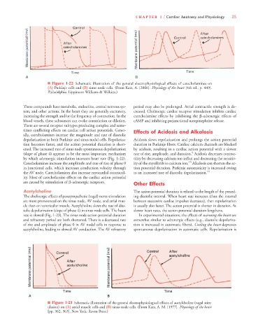
■ Figure 1-22 Schematic illustration of the general electrophysiological effects of catecholamines on
(A) Purkinje cells and (B) sinus node cells. (From Katz, A. [2006]. Physiology of the heart [4th ed., p. 449].
Philadelphia: Lippincott Williams & Wilkins.)
These compounds have metabolic, endocrine, central nervous sys- period may also be prolonged. Atrial contractile strength is de-
tem, and other actions. In the heart they are generally excitatory, creased. Cholinergic cardiac receptor stimulation inhibits cardiac
increasing the strength and/or the frequency of contraction. In the catecholamine effects by inhibiting the -adrenergic effects of
blood vessels, these substances can evoke constriction or dilation. cAMP and inhibiting prejunctional norepinephrine release.
There are several receptor subtypes producing complex and some-
times conflicting effects on cardiac cell action potentials. Gener- Effects of Acidosis and Alkalosis
ally, catecholamines increase the magnitude and rate of diastolic
depolarization in both Purkinje and sinus nodal cells. Repolariza- Acidosis slows repolarization and prolongs the action potential
tion becomes faster, and the action potential duration is short- duration in Purkinje fibers. Cardiac calcium channels are blocked
ened. The increased rate of sinus node spontaneous depolarization by acidosis, resulting in a cardiac action potential with a slower
5
(slope of phase 4) appears to be the most important mechanism rate of rise, amplitude, and duration. Acidosis decreases contrac-
by which adrenergic stimulation increases heart rate (Fig. 1-22). tility by decreasing calcium ion influx and decreasing the sensitiv-
45
Catecholamines increase the amplitude and rate of rise of phase 0 ity of the myofibrils to calcium ion. Alkalosis can shorten the ac-
in junctional cells, which increases conduction velocity through tion potential duration. Purkinje automaticity is increased owing
the AV node. Catecholamines also increase myocardial contractil- to an increased rate of diastolic depolarization. 44
ity. Most of catecholamine effects on the cardiac action potential
are caused by stimulation of -adrenergic receptors. Other Effects
Acetylcholine The action potential duration is related to the length of the preced-
The cholinergic effects of parasympathetic (vagal) nerve stimulation ing diastolic interval. When heart rate increases (thus the interval
are more pronounced on the sinus node, AV node, and atrial mus- between successive cardiac impulses decreases), then repolarization
cle than on ventricular muscle. Acetylcholine slows the rate of dias- is usually also faster. The action potential is shorter in duration. At
tolic depolarization (slope of phase 4) in sinus node cells. The heart slower heart rates, the action potential duration lengthens.
rate is slowed (Fig. 1-23). The sinus node action potential duration In experimental situations, the effects of warming the heart are
and refractory period are both shortened. There is a decreased rate somewhat similar to adrenergic effects (e.g., diastolic depolariza-
of rise and amplitude of phase 0 in AV nodal cells in response to tion is increased in automatic fibers). Cooling the heart depresses
acetylcholine, leading to slowed AV conduction. The AV refractory spontaneous depolarization in automatic cells. Repolarization is
■ Figure 1-23 Schematic illustration of the general electrophysiological effects of acetylcholine (vagal stim-
ulation) on (A) atrial muscle cells and (B) sinus node cells. (From Katz, A. M. [1977]. Physiology of the heart
[pp. 362, 363]. New York: Raven Press.)