
Page 53 - Cardiac Nursing
P. 53
92806_c01.qxd 11/21/11 10:30 AM Page 29
CHAPTER 1 / Cardiac Anatomy and Physiology 29
Calcium ion, the initiator and regulator of contraction, is the Molecular Basis for Contraction
major link between excitation and contraction. The intracellular
calcium concentration is directly and indirectly influenced by the The thick filaments are composed primarily of the protein myosin.
amount of calcium transported in and out of the cell across the Myosin is large, consisting of six subunits: two heavy chains and
sarcolemma. 54 Calcium sarcolemmal fluxes are affected by the four light chains per molecule. The two heavy chain subunits are
membrane potential and by sodium and potassium ion concen- coiled to form a long, rod-like tail at one end. At the opposite end
trations and transcellular fluxes. Conversely, potassium flux of the long myosin heavy chain, a head protrudes from each sub-
through the calcium-regulated potassium channel and sodium unit. Groups of myosin tails are arranged to form the rigid back-
flux during sodium–calcium exchange are affected by the intracel- bone of the thick filament. The heads are the site of ATP break-
lular concentration of calcium ion. down and interaction with the thin filaments. Heads project
outward in a spiral along the length of the thick filament. At the
center of the filament, the molecules reverse direction, leaving a
MECHANICAL CHARACTERISTICS bare region from which no heads protrude. The small light chains
OF CARDIAC CELLS are nestled in the angle between head and tail, two per heavy
chain. Both heavy and light chains are members of multigene
Overview of Contraction families and exist in several forms, called isoforms. Variation in iso-
form composition may modify the rate or intensity of myosin
As seen in Figure 1-15, the myofibril is composed of a series of re- chemical activity, which may modify the contractile properties of
peating units, called sarcomeres. Sarcomeres are the basic func- the tissue. Age, mechanical loading, or metabolic or hormonal
tional and structural units of the myofibril. Dark-staining Z lines state may modify isoform composition.
mark the ends of the sarcomere. Attached to the Z line are the The thin filaments are composed of bead-shaped molecules of
thin filaments. The center of the sarcomere is composed of the the protein actin arranged in an intercoiled, double-stranded
dark-appearing thick filaments. Interdigitating thin and thick fil- chain. Two other proteins, troponin and tropomyosin, are located
aments overlap to a variable extent. Shortening alters the amount on the thin filaments at periodic intervals (Fig. 1-27). Actin inter-
of thick and thin filament overlap: filament proteins interact caus- acts with the thick-filament protein, myosin, resulting in the trans-
ing the filaments slide past one another. duction of the chemical energy of ATP into mechanical energy.
The individual thick and thin filaments do not themselves Troponin and tropomyosin are called regulatory proteins because
change in length; the sarcomere (and the muscle as a whole) short- they modify the interaction of actin and myosin (Figs. 1-28, 1-29).
ens. If shortening of the sarcomere (or the muscle cell) is pre- Myosin is an enzyme that breaks down the high-energy ATP
vented, the interaction of thick and thin filaments is manifested as molecule. During the resting state, the products of ATP break-
tension or force generation. Such a contraction is termed isometric. down remain bound to the myosin head. When myosin interacts
When a stimulated muscle is allowed to shorten, tension is not in- with actin, the rate of ATP turnover is greatly increased. The
creased, and the contraction is said to be isotonic (Fig. 1-26). In chemical energy released from ATP is converted to the mechani-
the heart, early systolic contraction is primarily isometric, that is, cal energy of contraction and heat.
tension increases and muscle length remains fairly constant. Later According to the cross-bridge theory, a bond or crossbridge
in systole, the contraction is primarily isotonic, that is, the heart forms during muscle contraction, linking thick and thin filaments.
muscle shortens and the blood is expelled into the aorta, whereas The protuberant myosin head contains an actin-binding site and
little additional tension is developed. forms the crossbridge. This crossbridge is capable of binding,
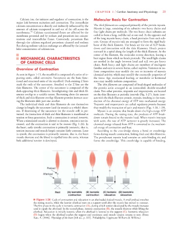
■ Figure 1-26 Cycle of contraction and relaxation in an afterloaded skeletal muscle. A small preload stretches
the resting muscle, while the heavier afterload rests on a support until after the muscle has started to contract.
The first phase in the cycle is isometric contraction (A), during which tension developed by the muscle increases
until it equals the afterload. In the second phase, isotonic contraction (B), the muscle shortens while lifting the
afterload. Relaxation is initially isotonic when the afterload is lowered to the support (C). Isometric relaxation
(D) begins when the afterload reaches the support and continues until muscle tension returns to zero. (From
Katz, A. [2006]. Physiology of the heart [4th ed., p. 322]. Philadelphia: Lippincott Williams & Wilkins.)