
Page 51 - Cardiac Nursing
P. 51
92806_c01.qxd 11/21/11 10:30 AM Page 27
CHAPTER 1 / Cardiac Anatomy and Physiology 27
Ventricular Conduction Purkinje fibers are sparsely distributed in the basal (upper) sec-
tions of the ventricles and septum, particularly in the right ventri-
The excitation impulse travels quickly through the His-Purkinje cle and the septum. The basal and posterior portions of both ven-
system. The His-Purkinje cells have the most rapid conduction ve- tricles and the basal interventricular septum are the last areas to be
locities in the heart, approximately 1.5 to 2 m/s in the His bun- activated, at approximately 80 milliseconds. 49
44
dle and 2 to 4 m/s in the Purkinje system. The cardiac impulse Although Purkinje fibers conduct the cardiac impulse more
next spreads rapidly (approximately 0.08 seconds), in a sequential rapidly than other cardiac cells, Purkinje cells in the distal termi-
manner from the common His bundle through the bundle nations of the conducting system have longer action potential du-
branches, then through the extensive ramifications of the Purkinje rations and refractory periods than do ventricular muscle fibers
fiber system, and finally through ventricular muscle. Ventricular (see previous). Because conduction is slower in cells with longer
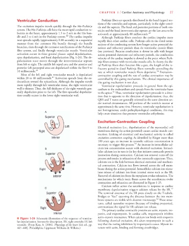
activation occurs in three general phases: septal depolarization, action potential durations and refractory periods, the conduction
apex depolarization, and basal depolarization (Fig. 1-24). The de- velocity of the cardiac impulse is slowed at the point where Purk-
polarization wave moves through the interventricular septum inje fibers connect with ventricular muscle cells. In theory, the dis-
from left to right. The middle left septal area and the anterior and tal Purkinje fibers then function like a gate, the length of the re-
posterior left paraseptal areas are depolarized within the first 0 to fractory period in distal Purkinje fibers normally controlling the
10 milliseconds. 49 rate at which ventricular muscle fibers depolarize. 50 Excitation–
Most of the left and right ventricular muscle is depolarized contraction coupling and the rate of cardiac contraction may be
within 20 to 40 milliseconds. 49 Activation spreads from the en- controlled by this gating mechanism. The clinical importance of
docardium toward the epicardium. Although the impulse travels this gating mechanism is not clear.
more rapidly through left ventricular tissue, the right ventricular Ventricular repolarization proceeds in general from the epi-
wall is thinner. Thus, the full thickness of the right ventricle gen- cardium to the endocardium and spreads from the ventricular bases
erally depolarizes prior to the left. The first epicardial depolariza- to the apices. Thus, ventricular repolarization proceeds in a direc-
51
tion usually occurs in the lower right ventricular wall. tion that is opposite to the direction of depolarization; thus, the
QRS and T waves are generally oriented in the same direction un-
der normal circumstances. All portions of the ventricle recover at
approximately the same time. However, ventricular repolarization is
not homogeneous; under pathophysiological conditions, this may
help create situations that promote ventricular arrhythmias.
Excitation–Contraction Coupling
Electrical excitation (i.e., depolarization of the myocardial cell
membrane during the action potential) causes cardiac muscle con-
traction. Linking of electrical and mechanical activity is called
excitation–contraction coupling. As identified by Ringer more than
100 years ago, an increase in cytosolic calcium concentration is
necessary to trigger this process. 52 An increase in intracellular cal-
cium ion concentration occurs with electrical excitation. Intracel-
lular calcium ion in turn is the key that initiates contractile protein
interaction during contraction. Calcium ion removal turns off the
process and results in relaxation of the contractile apparatus. Thus,
calcium ion is the link between electrical excitation and mechani-
cal contraction. Calcium ion flows inward across the cell mem-
brane during the action potential. Intracellular calcium ion stimu-
lates release of calcium ion from internal stores such as the SR.
Removal of calcium ion from the myoplasm evokes relaxation. The
mechanisms by which ionic fluxes across the sarcolemma evoke
contraction and relaxation are illustrated in Figure 1-25.
Calcium influx across the sarcolemma in response to cardiac
membrane depolarization triggers calcium release by the SR. 17
The terminal cisternae of the SR press closely on the T-tubule.
Bridges or “feet” spanning the distance between the two mem-
53
brane systems are visible with electron microscopy. These struc-
tures, called ryanodine receptors (because of binding properties),
communicate the signal for SR calcium ion release.
The primary cardiac contractile proteins are actin, myosin, tro-
ponin, and tropomyosin. In cardiac cells, tropomyosin inhibits
actin–myosin interaction. When calcium ion binds with troponin
■ Figure 1-24 Schematic illustration of the sequence of ventricu-
lar depolarization. See text for description. RV, right ventricle; LV, left following electrical excitation, this alters tropomyosin in such a
ventricle. (From Katz, A. [2006]. Physiology of the heart [4th ed., pp. way that the resting inhibition by tropomyosin ceases. Myosin in-
447–448]. Philadelphia: Lippincott Williams & Wilkins.) teracts with actin, binding and forming crossbridges.