
Page 50 - Cardiac Nursing
P. 50
92806_c01.qxd 11/21/11 10:30 AM Page 26
26 PA R T I / Anatomy and Physiology
delayed, and conduction is decreased. Arrhythmias may occur node normally initiates the electrical impulse that is then con-
during cooling, which is clinically relevant for the cardiac surgical ducted to other areas of the myocardium, depolarizing other cells
patient who has been subjected to hypothermia and for the pa- of the conducting system before those cells have time to sponta-
tient experiencing hypothermia caused by exposure. neously depolarize to threshold. The electrical impulse appears to
Stretching cardiac fibers increases the rate of diastolic depolar- spread outward in relatively concentric circles from the sinus node
ization and makes the maximal diastolic potential less negative in through the atria, moving in approximately 0.1 second from the
automatic fibers. Myocardial fiber stretch may cause arrhythmias upper right atrium to the posterior left atrium. Conduction ve-
during heart failure. locity (speed with which the impulse spreads) through the atria is
approximately 0.8 to 1 m/s (Table 1-5). Conduction velocities are
not equal through the atria; conduction is more rapid by way of
PROPAGATION OF THE the Bachmann bundle into the left atrium than in other areas of
CARDIAC IMPULSE the interatrial septum. There are specialized conduction pathways
in the atrium as in the ventricle, but the functional significance of
The spread of the cardiac impulse through the heart depends upon the atrial fibers is less clear. Generally, the impulse travels radially
several factors, including (1) anatomic characteristics of the con- within the atria. Atrial repolarization spreads in the same direction
ducting system, (2) structural characteristics of cells (e.g., cardiac as depolarization.
cell type and diameter, arrangement of low-resistance intercalated
discs, and contiguity to other cells capable of conducting current), Junctional Conduction
and (3) electrophysiological state of the cell membrane (i.e., rest-
ing and threshold potentials, ionic concentrations and conduc- The cardiac impulse is not conducted through the connective tis-
tances, rate and magnitude of depolarization and repolarization, sue of the cardiac skeleton, so cardiac muscle tissue in the AV
duration of the action potential and the refractory period). As in a junction provides the only pathway for electrical conduction from
battery, there is energy stored across the cell membrane. When one the atria to the ventricles. Conduction velocity through the AV
segment of the membrane depolarizes, positive charge enters the node is approximately 0.05 m/s, although in some areas it has
cell, and an electrical circuit is established along the cell. 5 been found to be as slow as 0.02 m/s.
In general, current flows more easily inside the cell and to ad- The rate of impulse conduction through the AV junction is in-
jacent cells across the intercalated discs at tight junctions than lat- fluenced by the atrial site at which the impulse enters the junc-
47
erally across adjacent, highly resistant areas of cell membranes. If tional area. An initial normal slowing of conduction through the
the current is sufficient to depolarize adjacent cells, a wave of de- AV junction with a later increase in the speed of conduction is cor-
polarization is propagated and spreads rapidly from cell to cell. related with electrophysiological differences in atrionodal, nodal,
48
Thus, the cardiac tissue behaves essentially as a syncytium, al- and nodal-His cells. Other mechanisms have been postulated for
though propagation may be somewhat discontinuous. 46 the slowing of conduction through the junction, including the
As the impulse spreads through the heart, it depolarizes tissue small size of the junctional conducting cells and the amounts of
that has recovered and is excitable, but it cannot depolarize tissue connective tissue interspersed among conducting cells.
that is still refractory. Because the cardiac impulse spreads rapidly The term decremental conduction describes the condition
through the atria, slowly through the AV junction, and then rapidly when a propagating impulse becoming successively weaker. The
through the ventricles, both atria contract almost synchronously, extent that decremental conduction normally occurs in the AV
the ventricles have time to receive blood from the contracting atria, junction is debatable. Decremental conduction can lead to AV
and then both ventricles contract almost synchronously. blocks. Slowing of the cardiac impulse at the AV junction pre-
vents the atria and ventricles from contracting simultaneously
Atrial Conduction and protects the ventricles from the abnormally fast heart rates
that can be generated in the atria under abnormal situations. Pre-
Sinus node cells normally have the fastest rate of spontaneous de- excitation syndromes are evoked when there are accessory junc-
polarization and thus set the pace of cardiac excitation. The sinus tional pathways. 27
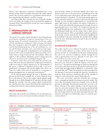
Table 1-5 ■ NORMAL CARDIAC ACTIVATION SEQUENCE
Normal Sequence Conduction Time for Impulse to Traverse Rate of Automatic
of Activation Velocity (m/s) Structure (in seconds) Discharge (per minute)
Sinoatrial node — 60–100
Atrial myocardium 0.8–1 r 0.15 None
AV node 0.02–0.05 See text
AV bundle 1.2–2 s 40–55
Bundle branches 1.5–2 0.08
Purkinje network 2–4 25–40
r
Ventricular myocardium 0.3–1 0.08 None
AV, atrioventricular; m, meters; s, second; ~, approximately.
Adapted from Katz, A. M. (1977). Physiology of the heart (p. 259). New York: Raven Press.