
Page 490 - Cardiac Nursing
P. 490
66
10.
Apt
10.
ara
qxd
9/2
0
0-5
9/0
M
66
8 A
009
8:2
P
p46
P
e 4
g
K34
0-c
21_
K34
L L LWB K34 0-c 21_ p46 0-5 10. qxd 0 9/0 9/2 009 0 0 8:2 8 A M P a a g e 4 66 Apt ara
LWB
LWBK340-c21_21_p460-510.qxd 09/09/2009 08:28 AM Page 466 Aptara
466 P A R T III / Assessment of Heart Disease
for infection, and adequate collateral circulation. 65 The most
common insertion site is the radial artery due to the presence of Table 21-2 ■ RISK FACTORS AND ACTIONS TO PREVENT
collateral circulation, which decreases the risk of vascular compli- COMPLICATIONS ASSOCIATED WITH ARTERIAL
cations. The radial and ulnar artery/superficial palmar arteries CATHETERIZATION
provide a dual blood supply to the hand. Although the Allen test Risk Factors Preventive Actions
has traditionally been used to evaluate collateral circulation, rec-
ommendations in the literature regarding its use are equivo- • Catheter 20 gauge • Use of heparinized flush solution to
cal. 66,67 In addition, the implications of a positive Allen test are • Catheter in place 3 days maintain patency is equivocal.
equivocal. The positive findings may not be consistent with con- • Female Consideration should also be given
to risk of heparin-induced
• Low CO/hypotension
current Doppler ultrasonography evaluation nor does the pres- • Peripheral vascular disease thrombocytopenia. 72 77
ence of an abnormal Allen test reliably predict that the patient will • Vasopressor agents • Aspirate clot or discontinue line if
develop hand ischemia after radial artery cannulation. Given this • Anticoagulation (T risk) thrombosis is suspected
limited diagnostic accuracy, newer technologies including digit • Femoral (T risk) • Perform routine monitoring of distal
pressure measurement and plethysmography and Doppler ultra- • Systemic antithrombotics or perfusion (skin color, temperature and
capillary refill) and after line
anticoagulants (T risk)
sonography should be considered, particularly for patients who • Insertion site (femoral or manipulation
are at increased risk for complications from the catheterization axillary) • No beneficial effect from repeated flushes
(e.g., peripheral vascular disease or diabetes, previous extremity • Insertion site preparation • No effect from method of blood 78
sampling (waste versus nonwaste)
surgery or trauma, current anticoagulation or vasopressor therapy, • Catheter in place 5 7 days • Catheter length sheaths/arterial lines
• Insertion site
and hypotension). 68 (T risk)
The femoral artery is often an alternative to the radial artery. • Maintain system integrity
However, there is an increased risk of infection with the femoral • Monitor waveform for damping (may in-
site. The brachial artery is used less frequently because it does not dicate loose connections) See Table 21-1
have good collateral circulation, which in theory increases the
risk for diffuse distal ischemia. The axillary artery is a less com-
mon insertion site, with complication rates similar to radial and
femoral insertions. The dorsalis pedis artery is another option. flow just before aortic valve closure. Pressure in the aorta continues
Complication rates associated with the dorsalis pedis artery are to decrease and is reflected on the arterial pressure waveform as a
69
comparable to radial artery insertion. The dorsalis pedis should gradual downslope until the next ventricular systole. The interval
not be used if the patient has peripheral vascular disease or an ab- after the incisura when the aortic pressure continues to decrease is
sent posterior tibial pulse. In addition, the dorsalis pedis artery referred to as the diastolic run-off period, and the slope of this
pressures are higher than radial pressures, even in the supine po- period is affected by arterial stiffness and the rate at which the
sition. 70 79
blood flows into the periphery (vascular resistance).
The arterial pressure waveform changes its contour when
Complications Related to Arterial recorded at different sites along the arterial circuit 80 (Fig. 21-6).
Catheterization The pulse pressure and the systolic pressure increase, and the as-
cending limb of the waveform becomes steeper. In addition, the
The most common complication from arterial cannulation is incisura is gradually replaced by a later diastolic wave (dicrotic
temporary occlusion of the artery (radial 19.7%; femoral 1.5%), notch). The change in amplitude and contour of the arterial wave-
although permanent arterial occlusion is rare (0.09% and form is primarily caused by peripheral pulse wave reflection. 81
0.18%, respectively). 65 Given the risk and potentially severe
outcomes from arterial occlusion, distal perfusion (skin color,
temperature, and capillary refill) should be routinely assessed
postinsertion and any time the system is manipulated. 68,71
Bleeding is a rare complication for all insertion sites (0.6% to
1.6%), with increased incidence in femoral and axillary lines. 65 Systole Diastole
A summary of risk factors and actions to prevent complications
is presented in Table 21-2.
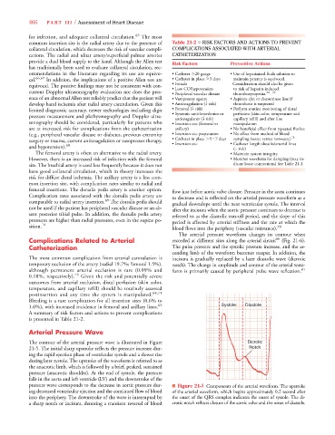
Arterial Pressure Wave
The contour of the arterial pressure wave is illustrated in Figure Dicrotic
21-5. The initial sharp upstroke reflects the pressure increase dur- Notch
ing the rapid ejection phase of ventricular systole and a slower rise
during later systole. The upstroke of the waveform is referred to as
the anacrotic limb, which is followed by a brief, peaked, sustained
pressure (anacrotic shoulder). At the end of systole, the pressure
falls in the aorta and left ventricle (LV) and the downstroke of the
pressure wave corresponds to the decrease in aortic pressure dur- ■ Figure 21-5 Components of the arterial waveform. The upstroke
ing decreased ventricular ejection and the continued flow of blood of the arterial waveform, which begins approximately 0.2 second after
into the periphery. The downstroke of the wave is interrupted by the onset of the QRS complex indicates the onset of systole. The di-
a sharp notch or incisura, denoting a transient reversal of blood crotic notch reflects closure of the aortic valve and the onset of diastole.