
Page 493 - Cardiac Nursing
P. 493
9/0
0
9/2
8:2
009
0-5
p46
10.
qxd
10.
69
e 4
69
ara
Apt
M
8 A
P
g
P
K34
K34
21_
0-c
LWBK340-c21_21_p460-510.qxd 09/09/2009 08:28 AM Page 469 Aptara
L L LWB K34 0-c 21_ p46 0-5 10. qxd 0 9/0 9/2 009 0 0 8:2 8 A M P a a g e 4 69 Apt ara
LWB
C HAPTER 2 1 / Hemodynamic Monitoring 469
CVP MONITORING or dorsal hand veins 120,121 or from a lower extremity insertion
site. 122 A key to the use of peripheral venous pressure (PVP) meas-
The CVP directly reflects right atrial pressure (RAP) and indi- urements is ensuring that there is continuity between the central
rectly reflects the preload of the right ventricle or RV end-diastolic and venous systems, which can be assessed by observing for an in-
pressure. The CVP is determinedby vascular tone, the volume of crease in the PVP with a sustained inspiratory effort (Valsalva) or
blood returning to the heart, the pumping ability of the heart, and the occlusion of the arm or leg above the catheter insertion site
122,123
patient position (supine, standing). (Fig. 21-9). 124 The PVP–CVP difference decreases with in-
The CVP is measured in the superior vena cava and the RAP creasing CVP, which may reflect vascular continuity. No sig-
is measuredfrom the proximal port of the PA catheter. The CVP nificant pressure differences were found on the basis of catheter
and RAP are generally similar as long as there is no vena caval ob- size (14 to 20 gauge) and patient position (as long as the system
120,122
struction. Normally, the CVP ranges from 3 to 8 cm H 2 O or 2 to was referenced to the phlebostatic axis) in patients who
125
6 mm Hg(1 mm Hg 1.36 cm H 2 O). In the supine/flat posi- were hemodynamically unstable, had a decreased ejection frac-
121
tion, a CVP ofless than 2 mm Hg may indicate hypovolemia, va- tion (EF), or were receiving vasoactive medications. In general,
sodilation, or increased myocardial contractility. An increased changes in CVP were mirrored by changes in PVP, which suggests
CVP may indicate increased circulatoryblood volume, vasocon- that trends in PVP may be useful. However, clinically significant
striction, or decreased myocardial contractility. An increased CVP PVP–CVP differences ( 2 to 3 mm Hg) may occur; thus, cau-
is also observed in RV failure, tricuspid insufficiency, positive- tion must be exercised when interpreting the absolute PVP val-
120,121,123–128
pressure breathing, pericardial tamponade, pulmonary embolus, ues.
and obstructive pulmonary disease. There is limited evidence that suggests that CVP measure-
ments can be obtained through an open-ended peripherally in-
serted central venous catheter (PICC). 129–131 Measurements from
Indications
the PICC, overestimate measurements from a central catheter by
The placement of a central venous or RA catheter is indicated to approximately 1 mm Hg and changes in the PICC CVP are
secure venous access, to administer vasoactive drugs and par- closely related to central line CVP measurements. 129,131 Accurate
enteral nutrition, and to monitor right heart preload. Hemody- PICC CVP measurements require correct positioning of the
namic monitoring using a CVP is most often performed when PICC tip (at the junction of the vena cava and RA). Passive hy-
cardiopulmonary function is relatively normal. Monitoring the drostatic pressure equilibration across the PICC line takes ap-
CVP has regained importance with the recognition of the effect of proximately 60 minutes, but this pressure gradient can be over-
right heart function on left heart function. 7 come immediately with a pressure line infusing fluid at 3 mL/h. 129
The CVP cannot be measured if the system has a valve (e.g.,
Groshong, PASV, or PowerPICC SOLO). A limitation of the use
Effect of Catheter Type and of peripheral versus central line during resuscitation is that the pe-
Location on CVP ripheral catheter cannot be used to obtain central venous oxygen
saturation, which is an end-point of resuscitation. 132
The CVP may be monitored via a central venous catheter or the
distal port of a multilumen catheter. 118 One study also suggests
that measurements obtained via tunneled catheters are compara- Limitations
ble to direct RA pressure measurements. 119
In cases where placement of a central venous catheter is not The CVP is not an accurate indicator of LV function or left heart
possible, recent studies suggest that the CVP can be indirectly preload. 133 In the presence of normal right heart function, severe
measured from a peripheral venous catheter inserted in the forearm deterioration of LV function may not be reflected by a change in
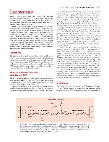
occlusion release
30 30
PVP
15 15
CVP
0 0
1 sec
■ Figure 21-9 Pressure waveforms from simultaneous PVP and CVP measurements demonstrating the ef-
fect of manual, circumferential, proximal arm occlusion followed by release. This increase indicates there is con-
tinuity between the central and peripheral venous systems. The PVP should not be used if this response is ab-
sent. (From Munis, J. R., Bhatia, S., Lozada, L. J. [2001]. Peripheral venous pressure as a hemodynamic variable
in neurosurgical patients. Anesthesia & Analgesia, 92, 174[.)