
Page 495 - Cardiac Nursing
P. 495
8:2
009
9/2
8 A
P
P
M
10.
0-5
p46
10.
9/0
0
qxd
g
K34
0-c
21_
K34
LWB K34 0-c 21_ p46 0-5 10. qxd 0 9/0 9/2 009 0 0 8:2 8 A M P a a g e 4 71 Apt ara
L L LWB
LWBK340-c21_21_p460-510.qxd 09/09/2009 08:28 AM Page 471 Aptara
71
71
e 4
Apt
ara
C HAPTER 2 1 / Hemodynamic Monitoring 471
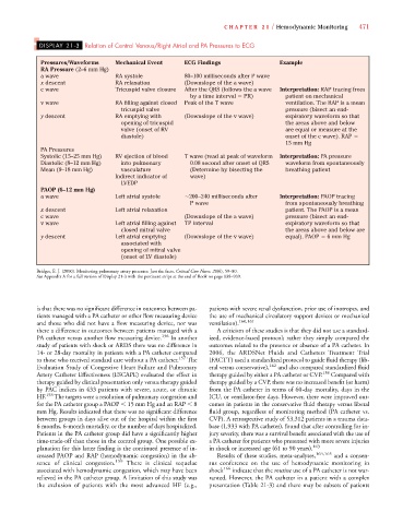
DISPLAY 21-3 Relation of Central Venous/Right Atrial and PA Pressures to ECG
Pressures/Waveforms Mechanical Event ECG Findings Example
RA Pressure (2–6 mm Hg)
a wave RA systole 80–100 milliseconds after P wave
x descent RA relaxation (Downslope of the a wave)
c wave Tricuspid valve closure After the QRS (follows the a wave Interpretation: RAP tracing from
by a time interval PR) patient on mechanical
v wave RA filling against closed Peak of the T wave ventilation. The RAP is a mean
tricuspid valve pressure (bisect an end-
y descent RA emptying with (Downslope of the v wave) expiratory waveform so that
opening of tricuspid the areas above and below
valve (onset of RV are equal or measure at the
diastole) onset of the c wave). RAP
15 mm Hg
PA Pressures
Systolic (15–25 mm Hg) RV ejection of blood T wave (read at peak of waveform Interpretation: PA pressure
Diastolic (8–12 mm Hg) into pulmonary 0.08 second after onset of QRS waveform from spontaneously
Mean (9–18 mm Hg) vasculature (Determine by bisecting the breathing patient
Indirect indicator of wave)
LVEDP
PAOP (6–12 mm Hg)
a wave Left atrial systole 200–240 milliseconds after Interpretation: PAOP tracing
P wave from spontaneously breathing
x descent Left atrial relaxation patient. The PAOP is a mean
c wave (Downslope of the a wave) pressure (bisect an end-
v wave Left atrial filling against TP interval expiratory waveform so that
closed mitral valve the areas above and below are
y descent Left atrial emptying (Downslope of the v wave) equal). PAOP 6 mm Hg
associated with
opening of mitral valve
(onset of LV diastole)
Bridges, E. J. (2000). Monitoring pulmonary artery pressures: Just the facts. Critical Care Nurse, 20(6), 59–80.
See Appendix A for a full version of Display 21-3 with the pertinent strips at the end of Book on page 938–939.
is that there was no significant difference in outcomes between pa- patients with severe renal dysfunction, prior use of inotropes, and
tients managed with a PA catheter or other flow measuring device the use of mechanical circulatory support devices or mechanical
and those who did not have a flow measuring device, nor was ventilation). 160,161
there a difference in outcomes between patients managed with a A criticism of these studies is that they did not use a standard-
PA catheter versus another flow measuring device. 156 In another ized, evidence-based protocol; rather they simply compared the
study of patients with shock or ARDS there was no difference in outcomes related to the presence or absence of a PA catheter. In
14- or 28-day mortality in patients with a PA catheter compared 2006, the ARDSNet Fluids and Catheters Treatment Trial
to those who received standard care without a PA catheter. 157 The (FACTT) used a standardized protocol to guide fluid therapy (lib-
Evaluation Study of Congestive Heart Failure and Pulmonary eral versus conservative), 162 and also compared standardized fluid
Artery Catheter Effectiveness (ESCAPE) evaluated the effect in therapy guided by either a PA catheter or CVP. 138 Compared with
therapy guided by clinical presentation only versus therapy guided therapy guided by a CVP, there was no increased benefit (or harm)
by PAC indices in 433 patients with severe, acute, or chronic from the PA catheter in terms of 60-day mortality, days in the
HF. 158 The targets were a resolution of pulmonary congestion and ICU, or ventilator-free days. However, there were improved out-
for the PA catheter group a PAOP 15 mm Hg and an RAP 8 comes in patients in the conservative fluid therapy versus liberal
mm Hg. Results indicated that there was no significant difference fluid group, regardless of monitoring method (PA catheter vs.
between groups in days alive out of the hospital within the first CVP). A retrospective study of 53,312 patients in a trauma data-
6 months, 6-month mortality, or the number of days hospitalized. base (1,933 with PA catheter), found that after controlling for in-
Patients in the PA catheter group did have a significantly higher jury severity, there was a survival benefit associated with the use of
time-trade-off than those in the control group. One possible ex- a PA catheter for patients who presented with more severe injuries
planation for this latter finding is the continued presence of in- in shock or increased age (61 to 90 years). 163
creased PAOP and RAP (hemodynamic congestion) in the ab- Results of these studies, meta-analyses, 164,165 and a consen-
sence of clinical congestion. 159 There is clinical sequelae sus conference on the use of hemodynamic monitoring in
associated with hemodynamic congestion, which may have been shock 166 indicate that the routine use of a PA catheter is not war-
relieved in the PA catheter group. A limitation of this study was ranted. However, the PA catheter in a patient with a complex
the exclusion of patients with the most advanced HF (e.g., presentation (Table 21-3) and there may be subsets of patients