
Page 491 - Cardiac Nursing
P. 491
8:2
009
9/2
P
M
8 A
9/0
10.
0-5
p46
0
qxd
10.
P
K34
K34
0-c
LWB K34 0-c 21_ p46 0-5 10. qxd 0 9/0 9/2 009 0 0 8:2 8 A M P a a g e 4 67 Apt ara
L L LWB
LWBK340-c21_21_p460-510.qxd 09/09/2009 08:28 AM Page 467 Aptara
21_
67
e 4
g
ara
Apt
67
C HAPTER 2 1 / Hemodynamic Monitoring 467
Interpretation of Arterial
Pressure Data
The mean arterial pressure (MAP), which represents the average
pressure through a cardiac cycle, is affected by the CO and sys-
temic vascular resistance (SVR) as described by the following
equation:
MAP CO SVR
■ Figure 21-6 Simultaneous recordings of aortic and radial arterial
pressure waves. (From Rowell, L. D., Brengelmann, G. L., Blackmon, Recall of the factors that affect SBP, DBP, and MAPs is important
R. J. et al. [1968]. Disparities between aortic and peripheral pulse when assessing changes in BP. The SBP is affected by left ventric-
pressures induced by upright exercise and vasomotor changes in man. ular stroke volume (SV), peak rate of ejection, and distensibility
7
7
Circulation, 37, 954–964.) of the vessel walls. The DBP is primarily affected by arterial pe-
ripheral resistance. The pulse pressure, which is the difference be-
tween systolic and diastolic pressures, is determined by SV, peak
rate of ventricular ejection, and the distensibility of the arterial
Reflection occurs when flow is impeded (i.e., when low-resistance ar- walls.
teries terminate in high-resistance vessels) and the pressure wave is On average, more central SBP (aortic, femoral, or brachial) is
reflected in a retrograde (backward) fashion. This retrograde pressure lower than radial SBP by 7 to 14 mm Hg and central DBP simi-
wave combines with the antegrade (forward) pressure pulse, and the lar to or higher than radial DBP by 1 to 9 mm Hg, while the MAP
arterial pressure is augmented. is unchanged and may be a more consistent value to evaluate and
The timing of the return of the reflected pressure wave from guide therapy. 88–90 The SBP differences change with aging (radial
the periphery is important because if the reflected wave arrives SBP aortic SBP), 91,92 vasoconstriction (radial brachial and
during systole it increases LV workload. 82,83 In young individuals, femoral), 86,87 vasodilation (femoral radial; aortic radial), 93,94
the reflected waves arrive at the heart after closure of the aortic and exercise (peripheral SBP may be as much as 80 mm Hg higher
valves, which beneficially augments the diastolic blood pressure than central aortic pressure). 80 Both peripheral wave reflection
(DBP) and thus coronary perfusion. However, with aging or in- and the end-pressure product, which is the result of the conver-
creased stiffness of the arteries (i.e., hypertension), the retrograde sion of kinetic energy from flowing blood into pressure as the
pulse wave arrives back at the heart during systole, which increases blood strikes the upstream-looking arterial catheter, cause aug-
the systolic blood pressure (SBP). 82,83 mentation of the peripheral SBP. Regardless of the source or site
95
Recognition of central aortic systolic pressure augmentation of BP measurement, a key point is that BP and perfusion are not
is important in evaluating the effects of various vasodilator synonymous, and a higher BP does not necessarily translate to
agents. Nitroglycerin and nitroprusside substantially decrease higher perfusion. 96
aortic pressure without a clinically measurable change in
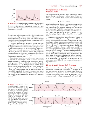
brachial pressure. 84,85 This effect, which is the result of the re- Direct Arterial Versus Cuff Pressure
duction in pulse–wave reflection (Fig. 21-7), may explain why
a patient may “look better” after the initiation of vasodilator There is no basis for the practice of comparing the intra-arterial
therapy even though there has been no marked decrease in pe- BP with the auscultatory or oscillometric BP to determine if
ripheral BP or preload. Conversely, vasoconstrictive agents one system should be followed to guide therapy. The direct
(e.g., norepinephrine) increase peripheral pulse pressure and method is based on pressure, whereas the oscillometric method
central aortic pressure, with femoral pressure higher than radial depends on flow-induced oscillations in the arterial wall. An er-
pressure. 86,87 roneous assumption is that pressure equals flow. As described by
NTG
Control NTG
■ Figure 21-7 Pressure wave 140 R R Ascending aorta
recorded directly in a central and pe- R
ripheral artery. Nitroglycerine 0.3 mg m mmHg
(SL) on average caused a fall of X X
11 mm Hg in aortic systolic pressure 70
more than the decrease in the brachial
systolic pressure. Note the effect on the
reflected (R) wave. (From Kelly, R. P., Brachial artery
Gibbs, H. H., O’Rourke, M. F., et al. R
[1990]. Nitroglycerine has more 140 R R
favourable effects on left ventricular af-
terload than apparent from measure- m mmHg
ment of pressure in a peripheral artery.
European Heart Journal, 11, 138–144.) 70
1 s 1 s