
Page 497 - Cardiac Nursing
P. 497
9/0
0
9/2
8:2
009
0-5
p46
10.
qxd
10.
73
e 4
73
ara
Apt
M
8 A
P
g
P
K34
0-c
21_
K34
LWB K34 0-c 21_ p46 0-5 10. qxd 0 9/0 9/2 009 0 0 8:2 8 A M P a a g e 4 73 Apt ara
L L LWB
LWBK340-c21_21_p460-510.qxd 09/09/2009 08:28 AM Page 473 Aptara
C HAPTER 2 1 / Hemodynamic Monitoring 473
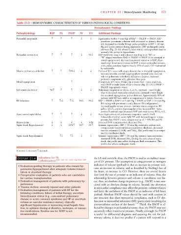
Table 21-3 ■ HEMODYNAMIC CHARACTERISTICS OF VARIOUS PATHOLOGICAL CONDITIONS
Hemodynamic Findings
Pathophysiology RAP PA PAOP SV CO Additional Findings
Pericardial tamponade ↑ ↑ ↑ ↓ ↓ Equalization (within 5 mm Hg) of RAP PAEDP PAOP; RAP
waveform: prominent x descent with attenuated or absent y descent
(d/t decreased ventricular filling); pulsus paradoxus (↓SBP 10 mm
Hg and ↓pulse pressure during inspiration; DBP unchanged); pulsus
alternans (Fig. 21-10); absent S 3 heart sound; cardiacpressures may be
normal if the patient is hypovolemic
Pericardial constriction ↑ ↑ ↑ ↓ N/↓ RAP waveform: steep x and y descent resulting in an “M”- or
“W”-shaped waveform; RAP PAEDP PAOP (if no tricuspid or
mitral regurgitation); decreased respiratory variation in RAP; Kuss-
maul’s sign (inspiratory increase in RAP in severe pericardial constric-
tion); pulsus paradoxus (approximately 33% of cases). CO maintained
by tachycardia
Massive pulmonary embolism ↑ ↑ ↑/N/↓ ↓ ↓ Increased RA v wave with steep y descent due to tricuspid regurgitation,
increased alveolar–arterial oxygen gradient (normal value does not
rule out pulmonary embolism), tachypnea, dyspnea, increased
pulmonic component of S 2 , pleuritic chest pain
Mitral regurgitation ↑ If amplitude of V wave 10 mm Hg or more than a wave amplitude,
read PAOP at nadir (base) of the x descent (Fig. 21-13C ); PAOP
PAEDP (regurgitant v wave)
Left ventricular failure N/↑ ↑ ↑ ↓ ↓ Pulmonary congestion or edema, S 3 or S 4 , increased a wave height
(due to decreased ventricular compliance); increased v wave height
due to mitral regurgitation, pulsus alternans. Approximately 50% of
patients with HF have mild or no impairment in systolic function
RV infarction ↑ ↑/↓ ↑/↓ N/↓ N/↓ RAP PAOP or RAP 1 to 5 mm Hg PAOP, or RAP 10 mm Hg,
RA tracing with prominent x and y descent (M configuration),
increased jugular venous pressure, systemic venous congestion, RV
gallop, split S 2 , positive hepatojugular reflux, increased RA a wave,
positive Kussmaul’s sign (increased RAP with inspiration), RV S 3 or S 4
Acute ventral septal defect ↑ ↑ ↑ ↓ ↓ Acute hypotension and pulmonary congestion, systolic thrill,
holosystolic murmur, acute right HF with increased jugular venous
pressure, late PAOP v wave, oxygen step up of 10% RA and PA
Hypovolemia ↓ ↓ ↓ ↓ ↓/N Increased SVR (compensatory), decreased Svo 2
Septic shock (hyperdynamic) ↓ ↓ ↓/N/↑ N/↑ N/↑ Systemic hypotension, SBP 90 mm Hg, metabolic acidosis with
compensatory hyperventilation (respiratory alkalosis), decreased
. This profile may be accompa-
vascular resistance (↓ SVR) and ↑Svo 2
nied by distributive shock.
Septic shock (hypodynamic) ↑↓ ↑↓ ↓ ↓ Systemic hypotension, SBP 90 mm Hg, systemic vasoconstriction
. During the early phase of septic
(increased SVR), decreased Svo 2
shock, this profile may reflect inadequate fluid resuscitation. This
profile also reflects cardiogenic shock.
N, normal; ↓, decreased; ↑, increased.
DISPLAY 21-4 Indications for PA the LA and ventricle; thus, the PAOP is used as an indirect meas-
Catheterization 158,160,161,163,164,166–169 ure of LV pressure. The assumption in using pressure as surrogate
indicator of volume (preload) is that an increase in pressure indi-
• Evaluation/guiding therapy in patients who remain hy-
potensive/hypoperfused after adequate volume resusci- cates an increase in volume, and as described by Starling’s law of
tation or standard therapy the heart, an increase in CO. However, there are several factors
• Preoperative evaluation of patients who are candidates that limit the use of pressure as an indicator of volume. First, the
for cardiac transplantation relationship between pressure and volume is curvilinear, not lin-
• Evaluation/management of patients with pulmonary hy- ear; thus, an absolute change in pressure (e.g., PAOP) is not asso-
pertension ciated with an absolute change in volume. Second, any alteration
• Trauma victims: severely injured and older patients in myocardial compliance may affect the pressure–volume relation
• Evaluation/management of patients with HF for the and limit the usefulness of the PAOP as an indicator of left heart
following conditions: failure of initial therapy, uncertain preload. Absolute PAOP values should be used with caution in
hemodynamic status (e.g., concomitant pulmonary
disease or acute coronary syndrome and HF or uncertain any situation that alters myocardial compliance, such as LV dys-
volume or vascular resistance status), clinically function or myocardial infarction (MI) (particularly involving the
174
significant hypotension or worsening renal/hepatic func- posteroinferior surface of the heart). Third, the PAOP is af-
tion, to optimize dosing of diuretics, inotropes, or vasoac- fected by changes in pericardial pressure; thus, the PAOP may not
tive medications. Routine use for ADHF is not accurately reflect transmural pressure. Finally, although the PAOP
recommended. is useful for differential diagnosis and assessing the risk for pul-
monary edema, it does not predict if a patient will respond to a