
Page 55 - Cardiac Nursing
P. 55
92806_c01.qxd 11/21/11 10:30 AM Page 31
CHAPTER 1 / Cardiac Anatomy and Physiology 31
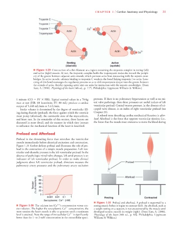
■ Figure 1-29 Cross section of a thin filament at a region containing the troponin complex in resting (left)
and active (right) muscle. At rest, the troponin complex holds the tropomyosin molecules toward the periph-
ery of the groove between adjacent actin strands, which prevents actin from interacting with the myosin cross-
bridges. In active muscle, calcium binding to troponin C weakens the bond linking troponin I to actin. Loos-
ening of this bond rearranges the regulatory proteins so as to shift tropomyosin deeper into the groove between
the strands of actin, thereby exposing active sites on actin for interaction with the myosin crossbridges. (From
Katz, A. [2006]. Physiology of the heart [4th ed., p. 117]. Philadelphia: Lippincott Williams & Wilkins.)
1 minute (CO SV HR). Typical normal values in a 70-kg pressure. If there is no pulmonary hypertension as well as no mi-
man at rest (HR: 68 beats/min; SV: 80 mL) produce a cardiac tral valve pathology, then these pressures are useful indices of left
output of 5,440 mL/min or 5.4 L/min. ventricular preload. Central venous pressure, in the absence of tri-
Stroke volume is determined by the degree of ventricular fill- cuspid valve disease, is an index of right ventricular preload (see
ing during diastole (preload), the force against which the ventricle Chapter 21).
must pump (afterload), the contractile state of the myocardium, A related term describing cardiac mechanical function is after-
and heart rate. In the remainder of this section, these factors are load. Afterload is the force that opposes ventricular ejection (i.e.,
discussed in more detail, and the manner in which they interact the forces that the muscle must overcome to move the blood during
to influence the mechanical function of the heart is described.
Preload and Afterload
Preload is the distending force that stretches the ventricular
muscle immediately before electrical excitation and contraction.
Figure 1-31 further defines preload and illustrates the role of pre-
load in the contraction of a simple muscle preparation. Left ven-
tricular end-diastolic pressure is the left ventricular preload. In the
absence of pathologic mitral valve changes, left atrial pressure is an
indicator of left ventricular preload. In order to make clinical
judgments about left ventricular preload, clinicians measure the
pulmonary artery pressures and the pulmonary artery occlusion
■ Figure 1-31 Preload and afterload. A preload is supported by a
■ Figure 1-30 The calcium ion (Ca 2 ) concentration versus ten- resting muscle before it begins to contract (left). An afterload, such as
sion relation. The higher the sarcoplasmic Ca 2 concentration, the a weight resting on a support, is not encountered by the muscle until
more tension the heart muscle is able to generate until a maximum developed tension exceeds its weight (right). (From Katz, A. [2006].
level is attained. Note the range of intracellular Ca 2 is significantly Physiology of the heart [4th ed., p. 83]. Philadelphia: Lippincott
lower than the 1- to 2-mM concentration in the extracellular space. Williams & Wilkins.)