
Page 504 - Cardiac Nursing
P. 504
LWBK340-c21_p460-510.qxd 09/09/2009 08:28 AM Page 480 Aptara
480 PA R T III / Assessment of Heart Disease
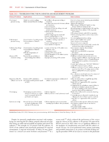
Table 21-5 ■ TROUBLESHOOTING THE PA CATHETER AND MEASUREMENT PROBLEMS
Clinical Problem Implications Possible Causes Interventions
Overdamped Falsely low systolic readings Air bubbles in the pressure tubing or Flush all air from system (including microbubbles).
pressure tracing Falsely increased diastolic readings transducer Remove excess stopcocks
More than three stopcocks between catheter Tighten all connections
and transducer Flush tubing of all blood (if unable to clear,
Loose connections change transducer-tubing set-up)
Collection of blood in tubing or in and Maintain pressure in infusion bag at 300 mm Hg
around transducer Aspirate blood from catheter if clot suspected
Catheter kinked internally or at (do not flush)
insertion site If PA catheter kinked, notify MD to reposition
Catheter wedged against vessel wall If fibrin occluding catheter, catheter may need to
Excessive tubing length ( 4 ft) be replaced
Clot or fibrin deposition on catheter tip Use noncompliant/wide-bore tubing
Underdamped Overestimation of systolic pressure Air bubbles in tubing, stopcocks, or Remove all air bubbles from system
pressure tracing Underestimation of diastolic transducer Limit tubing to 4 ft maximum
pressure Excessive tubing length ( 4 ft) Remove unnecessary stopcocks
Excess number of stopcocks If all attempts to resolve unsuccessful, consider the
addition of an in-line damping device
Catheter whip (fling) Overestimation of systolic pressure Location of distal tip of PA catheter near Assess dynamic response characteristics
or artifact Underestimation of diastolic pulmonic valve (troubleshoot system)
pressure Hyperdynamic heart Notify MD or qualified RN to reposition
Difficult interpretation of Looping of PA catheter in RV PA catheter
waveform External disruption of PA catheter system If fling fails to resolve, use mean pressure
Absence of PA Potential for air embolism or Balloon rupture If balloon is inflated without return of air into
f
occlusion tracing blood leaking from balloon Improper positioning of PA catheter syringe on passive deflation, assess for signs of air
A
port embolism (if present, place in Trendelenburg in
left lateral decubitus position, treat symptoms,
notify MD)
If stable, label balloon port “DO NOT WEDGE.”
Notify MD of need to replace catheter
If balloon is inflated to 1.5 mL, without change in
waveform from PA to PA wedge pattern, notify
MD or qualified RN of need to reposition
catheter
Once catheter is repositioned, assess the amount of
air required for wedge (ideal volume
1.25–1.5 mL)
Migration of the PA Presence of RV arrhythmias Accidental or spontaneous withdrawal of Inflate the balloon fully to engulf the tip of the
catheter into the RV Decreased diastolic pressure (equal catheter into the RV catheter and reduce ectopy
to RAP) Notify MD or, if approved for RN, reposition
catheter into PA
If compromised by arrhythmias, ensure balloon is
deflated and withdraw catheter into RA
(15–20 cm marking on PA catheter and RAP
waveform observed from distal port)
Overwedging Overwedging (eccentric balloon Catheter migration Slowly inflate balloon while constantly observing
inflation or inflation in a small Balloon position in small pulmonary vessel the waveform
vessel) is potential risk for PA If overwedge pattern observed, immediately stop
perforation and rupture inflation and allow balloon to deflate passively
Notify MD or, if approved for RN, reposition
catheter
Spontaneous wedge Potential for loss of blood supply Catheter migration (patient movement, Turn patient to side opposite catheter placement.
to branch of pulmonary vessel warming up of catheter after placement) Have patient straighten arm or turn head to
and risk of PA infarction dislodge catheter
Have patient gently cough
Notify MD or RN, reposition catheter
4
4
Modified from Gardner, P. E. (1993). Pulmonary artery pressure monitoring. AACN Clinical Issues in Critical Care Nursing, 4, 98–119.
Despite the potential complications associated with manipu- recent study, 209 which evaluated the performance of the correct
lating and removing the PA catheter, properly educated and qual- steps for removal of PA catheters in 60 patients (30 removed by
ified nurses can safely and successfully manipulate and remove PA RN/30 removed by MD), there was no significant difference in
catheters. 208 For example, in 125 patients with PA catheters, 39 the incidence of complications associated with PA catheter re-
(31%) of the catheters were incorrectly positioned (35 required moval. Of note, the nurses performed correct patient positioning
advancement, 4 required withdrawal), of which 36 were reposi- and provided instructions to the patient on breathe holding dur-
tioned by a critical care nurse (without complication). 206 In a ing the procedure 100% of the time in contrast to the physicians