
Page 503 - Cardiac Nursing
P. 503
qxd
79
79
9/2
9/0
0
e 4
0-5
g
P
p46
10.
10.
009
K34
0-c
21_
K34
LWB K34 0-c 21_ p46 0-5 10. qxd 0 9/0 9/2 009 0 0 8:2 8 A M P a a g e 4 79 Apt ara
L L LWB
LWBK340-c21_21_p460-510.qxd 09/09/2009 08:28 AM Page 479 Aptara
Apt
8:2
8 A
P
M
ara
C HAPTER 2 1 / Hemodynamic Monitoring 479
Pre-relaxation DISPLAY 21-6 Removal of PA Catheter
30 Mid-Point End-Expiration 1. Verify the order to remove the catheter
20
2. Assemble necessary equipment
Pulmonary Artery Occlusion Pressure (mm Hg) 10 0 Post-relaxation Ventilator 3. Document on the flow sheet the ECG rhythm and vital
Inspiratory Nadir
signs before initiating the procedure
4. Explain the procedure to the patient
5. Transfer IV infusions from PA catheter ports to side
30
port of introducer or discontinue IV solutions if appro-
priate
20
6. Ensure that the patient remains in hemodynamically
End-Expiration
stable condition after transfer of infusions to side port
10
7. Turn off any remaining infusions to distal and proximal
0
1 sec ports
8. Ensure that the balloon is deflated by lining up the red
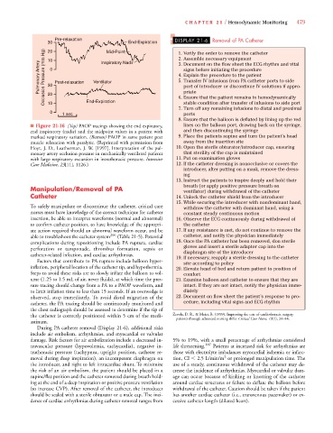
■ Figure 21-16 (Top) PAOP tracings showing the end expiratory, lines on the balloon port, drawing back on the syringe,
end inspiratory (nadir) and the midpoint values in a patient with and then discontinuing the syringe
marked respiratory variation. (Bottom) PAOP in same patient post 9. Place the patients supine and turn the patient’s head
muscle relaxation with paralytic. (Reprinted with permission from away from the insertion site
Hoyt, J. D., Leatherman, J. W. [1997]. Interpretation of the pul- 10. Open the sterile obturator/introducer cap, ensuring
monary artery occlusion pressure in mechanically ventilated patients that sterility of the cap is maintained
with large respiratory excursion in intrathoracic pressure. Intensive 11. Put on examination gloves
Care Medicine, 23[11], 1126.) 12. If the catheter dressing is nonocclusive or covers the
introducer, after putting on a mask, remove the dress-
ing
13. Instruct the patients to inspire deeply and hold their
breath (or apply positive pressure breath on
Manipulation/Removal of PA ventilator) during withdrawal of the catheter
Catheter 14. Unlock the catheter shield from the introducer
15. While securing the introducer with nondominant hand,
To safely manipulate or discontinue the catheter, critical care withdraw the catheter with dominant hand, using a
nurses must have knowledge of the correct technique for catheter constant steady continuous motion
insertion, be able to interpret waveforms (normal and abnormal) 16. Observe the ECG continuously during withdrawal of
to confirm catheter position, to have knowledge of the appropri- the catheter
ate action required should an abnormal waveform occur, and be 17. If any resistance is met, do not continue to remove the
able to troubleshoot the catheter system 206 (Table 21-5). Potential catheter, and notify the physician immediately
complications during repositioning include PA rupture, cardiac 18. Once the PA catheter has been removed, don sterile
perforation or tamponade, thrombus formation, sepsis or gloves and insert a sterile adaptor cap into the
diaphragm site of the introducer
catheter-related infection, and cardiac arrhythmias. 19.If necessary, reapply a sterile dressing to the catheter
Factors that contribute to PA rupture include balloon hyper- site according to policy
inflation, peripheral location of the catheter tip, and hypothermia. 20. Elevate head of bed and return patient to position of
Steps to avoid these risks are to slowly inflate the balloon to vol- comfort
ume (1.25 to 1.5 mL of air, never fluids), at which time the pres- 21. Examine balloon and catheter to ensure that they are
sure tracing should change from a PA to a PAOP waveform, and intact. If they are not intact, notify the physician imme-
to limit inflation time to less than 15 seconds. If an overwedge is diately
observed, stop immediately. To avoid distal migration of the 22. Document on flow sheet the patient’s response to pro-
catheter, the PA tracing should be continuously monitored and cedure, including vital signs and ECG rhythm
the chest radiograph should be assessed to determine if the tip of
the catheter is correctly positioned within 5 cm of the medi- Zevola, D. R., & Maier, B. (1999). Improving the care of cardiothoracic surgery
patients through advanced nursing skills. Critical Care Nurse, 19(1), 34–44.
astinum.
During PA catheter removal (Display 21-6), additional risks
include air embolism, arrhythmias, and myocardial or valvular
damage. Risk factors for air embolization include a decreased in- 5% to 19%, with a small percentage of arrhythmias considered
travascular pressure (hypovolemia, tachycardia), negative in- life threatening. 207 Patients at increased risk for arrhythmias are
trathoracic pressure (tachypnea, upright position, catheter re- those with electrolyte imbalances myocardial ischemia or infarc-
2
moval during deep inspiration), an incompetent diaphragm on tion, CI 2.5 L/min/m or prolonged manipulation time. The
the introducer, and right to left intracardiac shunt. To minimize use of a steady, continuous withdrawal of the catheter may de-
the risk of an air embolism, the patient should be placed in a crease the incidence of arrhythmias. Myocardial or valvular dam-
supine/flat position and the catheter removed during breath hold- age can occur because of kinking or knotting of the catheter
ing at the end of a deep inspiration or positive pressure ventilation around cardiac structures or failure to deflate the balloon before
(to increase CVP). After removal of the catheter, the introducer withdrawal of the catheter. Caution should be taken if the patient
should be sealed with a sterile obturator or a male cap. The inci- has another cardiac catheter (i.e., transvenous pacemaker) or ex-
dence of cardiac arrhythmias during catheter removal ranges from cessive catheter length (dilated heart).