
Page 70 - Cardiac Nursing
P. 70
LWBK340-c02_p042-068.qxd 06/30/2009 15:33 Page 46 Aptara
46 PA R T I / Anatomy and Physiology
Nitric Oxide. NO is a gas with an extremely short half-life
LOCAL REGULATION (seconds) that diffuses into vascular smooth muscle cells and
causes vasodilation. 40–42 NO production is stimulated by the en-
In addition to the systemic factors that affect vascular resistance, zyme nitric oxide synthase (NOS). There are two constitutive
there are local factors that control resistance. These factors include forms of NOS: endothelial NOS (eNOS) and neurological NOS
autacoids, endothelium-derived vasoactive substances, local meta- (nNOS). Inducible NOS (iNOS), which is present only under
bolic factors that match blood flow (oxygen transport) to metabo- pathological conditions, generates 100- to 1000-fold more NO
lism, autoregulation (see Chapter 3), and local heating and cooling. than the constitutive forms.
Shear stress and vasoactive substances are the primary factors
involved in the release of NO for control of vasomotor tone (Fig.
Autacoids
2-5). The activation of eNOS is different for these two mecha-
The autacoids (vasoactive substances) include histamine, sero- nisms. Shear stress through G proteins (Gs) leads to eNOS acti-
tonin, prostaglandin, and bradykinin. These factors most often vation, which via the inositol triphosphate (IP 3 ) pathway causes
compete with adrenergic (vasoconstrictive) effects and exert a lo- hyperpolarization of the endothelial cells, which allows calcium to
cal vasodilatory effect, which can improve tissue perfusion. The flow into the cell. 43,44 The increased intracellular calcium binds to
autacoids are not involved in systemic regulation of blood pressure calmodulin, which releases eNOS from the inhibitory protein
or total peripheral resistance; however, they initiate or modify the calveolin. The eNOS catalyzes the conversion of L-arginine to
vascular response to other stimuli. NO. After NO is formed in the endothelial cells, it diffuses out of
the endothelial cell to the vascular smooth muscle and as de-
scribed below causes vasodilation. Nitric oxide also has secondary
Endothelium-Derived Vasoactive vasodilatory effects through the inhibition of the release of the
Substances vasoconstrictor endothelin-1 (ET 1 ), although this beneficial effect
decreases with age. 45,46
The vascular endothelium, which is a single layer of squamous
cells in the tunica intima that lines the entire vascular tree, mod- Autocoids and hormones cause the release of NO from the en-
47
ulates vascular tone by secreting dilator and constrictor sub- dothelium (Fig. 2-6). These substances (Table 2-2) cause the re-
stances. In addition, the endothelium affects platelet adhesion and lease of IP 3 , which leads to an increase in intracellular calcium and
aggregation and under basal conditions substances secreted by the subsequently stimulates the release of NO. Additionally, NO de-
37
endothelium affect the clotting cascade. The endothelium is also creases sympathetic vasoconstriction by inhibiting the release of
48
involved in the regulation of vascular smooth muscle prolifera- norepinephrine at the supraspinal, spinal, and synaptic levels.
tion. 37 The proposed functions of the vascular endothelium Clinically, nitrovasodilators (e.g., nitroglycerin and sodium nitro-
(Table 2-1) require an intact endothelium. 38 The control of vas- prusside) cause vasodilation by the donation of NO or NO-like
49,50
cular tone involves cross talk between the vasodilators nitric oxide compound. Of note, nitroglycerin-induced coronary artery
(NO), prostaglandins, and endothelial-derived hyperpolarizing vasodilation does not require the presence of an intact endothe-
factor (EDHF) and the vasoconstrictors endothelin-1 and prosta- lium. In addition, ACE inhibitors, calcium channel blockers,
cyclin (Fig. 2-4). A discussion of each of these factors related to statins, and phosphodiesterase inhibitors indirectly stimulate NO
51
vascular control follows. A summary of the stimuli that cause the release or enhance its bioavailability.
release of each of the factors is presented in Table 2-2. Nitric oxide also inhibits platelet activation, aggregation, and
adhesion (i.e., anticoagulant/profibrinolytic phenotype), leukocyte
Endothelium-Derived Relaxing Factors adhesion, 52 vascular smooth muscle proliferation, and it inhibits
The seminal observation that endothelium is a key mediator of endothelial cell apoptosis but stimulates vascular smooth muscle
37,53
38
vascular reactivity was made in 1980. The ability of the artery to apoptosis (Fig. 2-6). With aging there is a decrease in NO pro-
relax was attributed to the elusive substance, EDRF, which was duction and increased endothelial cell apoptosis, which leads to a
later identified as NO. 39,40 Although NO is the major EDRF, decrease in the protective effect of NO against platelet aggregation
and vasoconstriction decreases. There is also diminished NO pro-
other relaxing factors such as prostacyclin [prostaglandin I 2
(PGI 2 )] EDHF are also produced. duction with disease processes (e.g., hypertension, diabetes, post-
myocardial infarction reperfusion injuries) and altered defense
mechanisms. 44,54 The loss of the protective endothelium, de-
creased NO production, and increased NO degradation may fos-
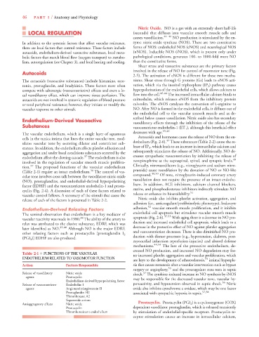
Table 2-1 ■ FUNCTIONS OF THE VASCULAR
ter increased platelet aggregation and vascular proliferation, which
ENDOTHELIUM RELATED TO VASOMOTOR FUNCTION 55
are keys to the development of atherosclerosis, intimal hyperpla-
Action Factors Responsible sia that causes restenosis after a vascular intervention such as bypass
surgery or angioplasty, 53 and the procoagulant state seen in septic
Release of vasodilatory Nitric oxide shock. The cytokine-induced increase in NO synthesis by iNOS
37
agents Prostacyclin
Endothelium-derived hyperpolarizing factor may be responsible for the decreased vascular tone, vascular hy-
56
Release of vasoconstrictor Endothelin-1 poreactivity, and hypotension observed in septic shock. Nitric
agents Angiotensin/angiotensin II oxide also inhibits cytochrome c oxidase, which may be one factor
Prostaglandin H2 associated with cytopathic hypoxia in sepsis. 57,58
Thromboxane A2
Superoxide anions
Antiaggregatory effects Nitric oxide Prostacyclin. Prostacyclin (PGI 2 ) is a cyclooxygenase (COX)
Prostacyclin dependent vasodilator prostaglandin, which is released transiently
Thromboresistant endothelium by stimulation of endothelial-specific receptors. Prostacyclin re-
ceptor stimulation causes an increase in intracellular calcium,