
Page 73 - Cardiac Nursing
P. 73
g
68.
2-0
g
68.
q
q
Pa
q
2-0
9 A
9 A
p
p
e 4
04
04
e 4
g
6/3
1
5:3
6/3
1
009
009
0/2
0/2
5:3
xd
xd
Pa
Pa
3
0
0
0
3
p
0-c
LWB
K34
02_
LWBK340-c02_
ta
LWB K34 0-c 02_ p p pp042-068.qxd 06/30/2009 15:33 Page 49 Aptara a a
ta
r
r
C HAPTER 2 / Systemic and Pulmonary Circulation and Oxygen Delivery 49
–
causes endothelium-dependent hyperpolarization, which in turn (TXA 2 ); superoxide anions (O 2 ); and components of the renin–
A
decreases cytosolic calcium and causes vasorelaxation through the angiotensin–aldosterone system. These substances are released in re-
various mediator-specific pathways (Fig. 2-8). 61 sponse to vasoconstrictive stimuli (see Fig. 2-4). Vasoconstriction
In hypertension or hypercholesterolemia, when NO-mediated also occurs as a result of a decrease in endothelial production of NO.
vasodilation is decreased, there may be a compensatory upregula-
tion of EDHF-mediated vasorelaxation. 64 Of note, there may be Endothelin-1. In humans there are three isoforms of en-
67
gender-specific EDHF compensatory response, with increased dothelin. ET 1 , which is the primary isoform in the cardiovascu-
EDHF activity in females but not in males. 62 However, oxidative lar system, is thought to be the most potent vasoconstrictor
stress associated with atherosclerosis, hyperhomocysteinemia, and known. ET 1 is an amino acid peptide that binds to vascular
possibly poorly controlled diabetes can decrease this compensa- smooth muscle membrane receptors ET A (located on vascular
tory response. 64–66 smooth muscle) and ET B (located on vascular smooth muscle and
endothelial surfaces). Binding of ET 1 to the ET A and ET B recep-
T
tors on the vascular smooth muscle activates the phospholipase C
Endothelium-Derived Contracting Factors
The endothelium-derived contracting factors include ET 1 , the vaso- (PLC)-IP 3 pathway, which increases intracellular calcium and the
constrictor prostanoids, PGH 2 , the precursor of thromboxane A2 phosphorylation of myosin kinase and causes prolonged muscle
contraction. In contrast, under normal resting conditions, the cir-
culating plasma level of ET 1 is very low and it acts locally, in a
paracrine fashion, to cause vasodilation through the endothelial
Ach, BK,SP τ synthesis of NO and PGI 2 . 68,69 The production of ET 1 can be
R augmented by shear stress, angiotensin II (AII), vasopressin, oxy-
gen free radicals, thrombin, and platelet-derived transforming
Endothelial
cells growth factor and inhibited by NO, atrial natriuretic polypeptide,
70
EDHF B-type natriuretic peptide, and prostacylin (Fig. 2-9).
2 2
The effects of ET 1 are important clinically. In pathological con-
1 Ca 3 Hyperpolarization
ditions such as heart failure, increased ET 1 levels are associated with
S
SK ca IK ca increased morbidity and mortalityy 71 and may play an important
64
Gap junctions role in the disease pathogenesis. For example, ET 1 stimulates the
Ga
K Hyperpolarization renin–angiotensin–aldosterone system, which enhances the conver-
EDHF
K K K Smooth sion of angiotensin I to AII, causing a synergistic augmentation of
70
muscle cells vasoconstriction and sodium retention. In addition, in heart fail-
68
ure, ET 1 has an negative inotropic effect. ET 1 may play a role in
salt-sensitive hypertension although the mechanism remains un-
BK ca Na /K K IR 68,72
ATPase clear. Unfortunately antagonism of ET 1 receptors has not been
found to improve outcomes for patients with heart failure or hy-
Hyperpolarization pertension. 73–75 ET 1 levels are increased with hypercholesterolemia
and increased ET 1 levels may be a marker for endothelial dysfunc-
tion. Possible pathological mechanisms for ET 1 in atherosclerosis
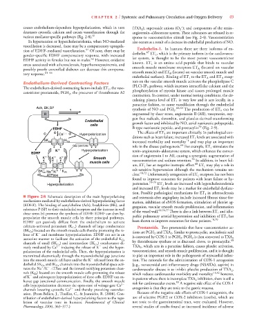
■ Figure 2-8 Schematic description of the main hyperpolarizing and restenosis after angioplasty include increased fibrous tissue for-
mechanisms mediated by endothelium-derived hyperpolarizing factor mation, inhibition of eNOS formation, stimulation of platelet ag-
(EDHF). The binding of acetylcholine (Ach), bradykinin (BK), and gregation, vascular smooth muscle proliferation, and inflammation
substance P (SP) to their endothelial receptors and the increase in wall of the vessel wall. 69,76,77 There is also a link between ET 1 and idio-
shear stress ( ) promote the synthesis of EDHF. EDHF can then hy-
perpolarize the smooth muscle cells by three principal pathways. pathic pulmonary arterial hypertension and inhibition of ET 1 has
78,79
EDHF can passively diffuse from the endothelium to activate been shown to improve outcomes for these patients.
calcium-activated potassium (K Ca ) channels of large conductance Prostanoids. Two prostanoids that have vasoconstrictive ac-
(BK Ca ) located on the smooth muscle cells thereby promoting the re- tions are PGH 2 and TXA 2 . Similar to prostacyclin, arachidonic acid
A
lease of K and membrane hyperpolarization. EDHF can act in an
A
is converted by COX-1 to PGH 2 . PGH 2 is then converted to TXA 2
autocrine manner to facilitate the activation of the endothelial K Ca by thromboxane synthase or as discussed above, to prostacyclin. 80
channels of small (SK Ca ) and intermediate (IK Ca ) conductance di-
A
rectly mediated by Ca 2
inducing the release of K and the hyper- TXA 2 , which acts in a paracrine fashion, causes platelet activation,
polarization of the endothelial cells. Then, the hyperpolarization is vasoconstriction, and smooth muscle proliferation, and it is thought
transmitted electronically through the myoendothelial gap junctions to play an important role in the pathogenesis of myocardial infarc-
into the smooth muscle cell layer and/or the K released from the en- tion. The rationale for the administration of COX-1 antagonists
dothelial SK Ca and IK Ca channels into the myoendothelial space acti- [e.g., nonsteroidal anti-inflammatory drugs (NSAIDs), aspirin] in
vates the Na /K ATPase and the inward rectifying potassium chan- cardiovascular disease is to inhibit platelet production of TXA 2 ,
nels (K IR ) located on the smooth muscle cells promoting the release which reduces cardiovascular morbidity and mortality; 81,82 however,
R R
of K and subsequent hyperpolarization of these cells. EDHF can en- in patients where there is incomplete TXA 2 inhibition, there is still a
A
hance gap junctional communication. Finally, the smooth muscle risk for cardiovascular events. A negative side effect of the COX-1
83
cells hyperpolarization decreases the open-state of voltage-gate Ca 2
channels lowering cytosolic Ca 2
and thereby provoking vasorelax- antagonists is that they are toxic to the gastric mucosa.
ation. (From Bellien, J., Thuillez, C., & Joannides, R. [2008]. Con- Because of the negative side effects of COX-1 antagonists, the
tribution of endothelium-derived hyperpolarizing factors to the regu- use of selective PGH2 or COX-2 inhibitors (coxibs), which are
lation of vascular tone in humans. Fundamental of Clinical not toxic to the gastrointestinal tract, were evaluated. However,
Pharmacology, 22(4), 363–377.) several studies of coxibs found an increased incidence of adverse