Page 76 - Cardiac Nursing
P. 76

3
                                        3
                                      5:3
                                         Pa
                                         Pa
                                         Pa
                                      5:3
                                  009
                                0/2
                                0/2
                                     1
                                     1
                                  009
                                           g
                                                 ta
                                                p
                                                p
                                                  r
                                                  r
                                                 ta
                                                p
                                            e 5
                                           g
                                           g
                                              2 A
                                              2 A
                                            e 5
                              6/3
                        68.
                          q
                      2-0
                        68.
                           xd
                           xd
                          q
                          q
                      2-0
         LWBK340-c02_
            K34
         LWB K34 0-c 02_ p p pp042-068.qxd  06/30/2009  15:33  Page 52 Aptara a a
         LWB
                    04
                    04
               0-c
                 02_
                              0
                              6/3
                             0
                             0
                  52    PA R T  I / Anatomy and Physiology
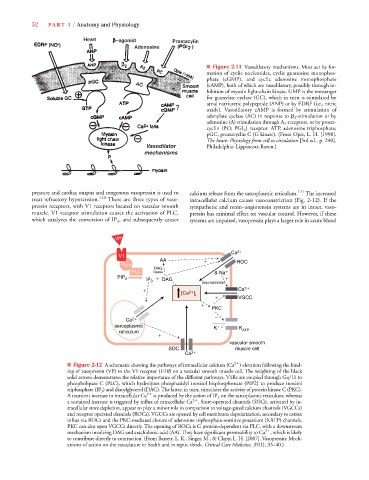
                                                                            ■ Figure 2-11 Vasodilatory mechanisms. Most act by for-
                                                                            mation of cyclic nucleotides, cyclic guanosine monophos-
                                                                            phate (cGMP), and cyclic adenosine monophosphate
                                                                            (cAMP), both of which are vasodilatory, possibly through in-
                                                                            hibition of myosin light-chain kinase. GMP is the messenger
                                                                            for guanylate cyclase (GC), which in turn is stimulated by
                                                                            atrial natriuretic polypeptide (ANP) or by EDRF (i.e., nitric
                                                                            oxide). Vasodilatory cAMP is formed by stimulation of
                                                                            adenylate cyclase (AC) in response to   2 -stimulation or by
                                                                            adenosine (A) stimulation through A 2 -receptors, or by prosta-
                                                                                                    A
                                                                            cyclin (PO; PGI 2 ) receptor. ATP, adenosine triphosphate;
                                                                            pGC, prostacyclin C (G kinase). (From Opie, L. H. [1998].
                                                                            The heart: Physiology from cell to circulation [3rd ed., p. 240].
                                                                            Philadelphia: Lippincott-Raven.)
                  pressure and cardiac output and exogenous vasopressin is used to  calcium release from the sarcoplasmic reticulum. 121 The increased
                  treat refractory hypotension. 120  There are three types of vaso-  intracellular calcium causes vasoconstriction (Fig. 2-12). If the
                  pressin receptors, with V1 receptors located on vascular smooth  sympathetic and renin–angiotensin systems are in intact, vaso-
                  muscle. V1 receptor stimulation causes the activation of PLC,  pressin has minimal effect on vascular control. However, if these
                  which catalyzes the conversion of IP 3 , and subsequently causes  systems are impaired, vasopressin plays a larger role in acute blood
                                              VP
                                                                                    Ca 2
                                               V1
                                                            AA            
          ROC
                                                G q/11
                                                          DAG
                                                   PLC    lipase              & Na
                                              PIP 2     IP 
  DAG
                                                         3
                                                                          depolarization
                                                       
              2
              Ca 2
                                                                   [Ca  ]
                                                                        i
                                                                              
       VGCC

                                                                             PKC

                                                 Ca 2
                                             sarcoplasmic                     K 
     K
                                               reticulum                               ATP

                                                                                   vascular smooth
                                                               SOC                   muscle cell
                                                                    Ca 2
                                                                                     2
                              ■ Figure 2-12 A schematic showing the pathways of intracellular calcium (Ca ) elevation following the bind-
                              ing of vasopressin (VP) to the V1 receptor (V1R) on a vascular smooth muscle cell. The weighting of the black
                                                              R
                                                              R
                              solid arrows demonstrates the relative importance of the different pathways. V1Rs are coupled through Gq/11 to
                              phospholipase C (PLC), which hydrolyzes phosphatidyl inositol bisphosphonate (PIP2) to produce inositol
                              triphosphate (IP 3 ) and diacylglycerol (DAG). The latter, in turn, stimulates the activity of protein kinase C (PKC).
                              A transient increase in intracellular Ca 2
  is produced by the action of IP 3 on the sarcoplasmic reticulum, whereas
                                                                       2
                              a sustained increase is triggered by influx of extracellular Ca . Store-operated channels (SOCs), activated by in-s
                              tracellular store depletion, appear to play a minor role in comparison to voltage-gated calcium channels (VGCCs)
                              and receptor operated channels (ROCs). VGCCs are opened by cell membrane depolarization, secondary to cation
                              influx via ROCs and the PKC-mediated closure of adenosine triphosphate-sensitive potassium (KATP) channels.
                              PKC can also open VGCCs directly. The opening of ROCs is G protein-dependent via PLC, with a downstream
                                                                                               2
                              mechanism involving DAG and arachidonic acid (AA). They have significant permeability to Ca , which is likely
                              to contribute directly to contraction. (From Barrett, L. K., Singer, M., & Clapp, L. H. [2007]. Vasopressin: Mech-
                              anisms of action on the vasculature in health and in septic shock. Critical Care Medicine, 35(1), 33–40.)
   71   72   73   74   75   76   77   78   79   80   81