
Page 78 - Cardiac Nursing
P. 78
r
ta
6/3
r
ta
p
4 A
p
p
q
xd
q
q
xd
0
6/3
0
0
4 A
3
3
5:3
5:3
Pa
g
g
Pa
Pa
g
0/2
e 5
e 5
0/2
1
1
009
009
68.
K34
p
p
K34
02_
02_
0-c
0-c
04
68.
2-0
2-0
04
LWBK340-c02_ pp042-068.qxd 06/30/2009 15:33 Page 54 Aptara a a
54 PA R T I / Anatomy and Physiology
dephosphorylation of myosin by the enzyme myosin light-chain crovascular blood flow and exchange, and postcapillary venous
phosphatase. The dephosphorylation facilitates the detachment of volume. 135,136 Although the radius of the capillaries is consider-
myosin from actin, resulting in relaxation. 130 ably smaller than the radius of the arterioles, the resistance is lower
The cytoplasmic calcium concentration is decreased through because of the increase in cross-sectional area.
uptake of calcium into the sarcoplasmic reticulum and transport
out of the cell across the plasma membrane by Ca 2
-ATPase ex- Volume Distribution
changer or a probable Na /Ca 2
exchanger. 34 Additionally, cal-
cium is decreased by closure of the membrane calcium channels At rest, the systemic veins contain as much as 60% to 80% of
through hyperpolarization 132 or pharmacologically with calcium the total blood volume, with 25% to 50% of this volume in the
channel blockers. small veins ( 1 mm in diameter). One-fourth of the total blood
volume is in the capacious splanchnic circulation. Although the
cross-sectional area is largest at the end of the capillaries, the
VOLUME AND FLOW largest volume of blood, as demonstrated in Figure 2-15, is in
DISTRIBUTION the venules because of the combination of cross-sectional area
and the length of the venules. 137 The remainder of the blood is
Resistance distributed in the aorta and systemic arteries (10%), the capil-
laries (5%), and the pulmonary bed and heart (15% to 25%). 2
The pressure decrease from the aorta to the small arteries is rela-
tively small, approximately 25 mm Hg (Fig. 2-14). 133 As much as Blood Flow
50% of the peripheral resistance appears to occur proximal to ves-
sels with diameters of 100 mm. This finding indicates the primary Definition of Flow
˙
sites for peripheral vascular resistance are the small arteries and the Blood flow (Q) is expressed in terms of volume of blood per unit
arterioles, although the exact location of the resistance vessels re- of time (volume/time). For example, the cardiac output, which is
mains equivocal. 134 defined as the liters of blood pumped out of the left ventricle into
Alterations in the diameter of the terminal arterioles or pre- the systemic circulation each minute, is usually expressed as liters
capillary blood vessels control capillary and venous pressures, mi- per minute.
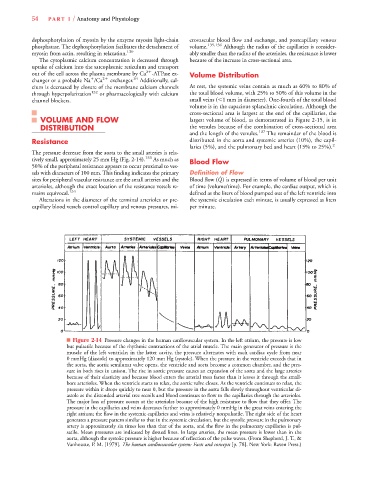
■ Figure 2-14 Pressure changes in the human cardiovascular system. In the left atrium, the pressure is low
but pulsatile because of the rhythmic contractions of the atrial muscle. The main generator of pressure is the
muscle of the left ventricle; in the latter cavity, the pressure alternates with each cardiac cycle from near
0 mmHg (diastole) to approximately 120 mm Hg (systole). When the pressure in the ventricle exceeds that in
the aorta, the aortic semilunar valve opens, the ventricle and aorta become a common chamber, and the pres-
sure in both rises in unison. The rise in aortic pressure causes an expansion of the aorta and the large arteries
because of their elasticity and because blood enters the arterial trees faster than it leaves it through the small-
bore arterioles. When the ventricle starts to relax, the aortic valve closes. As the ventricle continues to relax, the
pressure within it drops quickly to near 0, but the pressure in the aorta falls slowly throughout ventricular di-
astole as the distended arterial tree recoils and blood continues to flow to the capillaries through the arterioles.
The major loss of pressure occurs at the arterioles because of the high resistance to flow that they offer. The
pressure in the capillaries and veins decreases further to approximately 0 mmHg in the great veins entering the
right atrium; the flow in the systemic capillaries and veins is relatively nonpulsatile. The right side of the heart
generates a pressure pattern similar to that in the systemic circulation, but the systolic pressure in the pulmonary
artery is approximately six times less than that of the aorta, and the flow in the pulmonary capillaries is pul-
satile. Mean pressures are indicated by dotted lines. In large arteries, the mean pressure is lower than in the
aorta, although the systolic pressure is higher because of reflection of the pulse waves. (From Shepherd, J. T., &
Vanhoutte, P. M. [1979]. The human cardiovascular system: Facts and concepts [p. 78]. New York: Raven Press.)