
Page 872 - Cardiac Nursing
P. 872
60.
60.
009
0
2-8
9:4
9:4
2-8
p84
009
2
9
qx
d
9
6
2
/
0
37_
0-c
ar
a
K34
LWB
LWB K34 0-c 37_ p84 2-8 60. qx d 2 9 / 0 6 / / 2 009 0 9:4 0 P M Pa g e 8 48 Apt ar a
K34
LWBK340-c37_p842-860.qxd 29/06/2009 09:40 PM Page 848 Aptara
g
e 8
Pa
0 P
M
Apt
48
848 PA R T V / Health Promotion and Disease Prevention
patients with ischemia usually do not require such detailed obser-
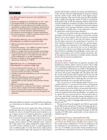
DISPLAY 37-4 General Guidelines for Risk Stratification vation. Patients with myocardial damage are limited by reduced
maximal cardiac output, which leads to early fatigue and pul-
Low Risk (all must be present to be classified at monary symptoms, rather than by chest pain. An effort should be
lowest risk) made to explain the symptoms related to CHF in such patients.
• Absence of significant LV dysfunction (i.e., EF 50%). In the patient with an MI, the symptoms could be caused by mi-
• Uncomplicated MI or revascularization procedure. tral valve insufficiency secondary to papillary muscle dysfunction,
• No resting or exercise-induced myocardial ischemia a dilated mitral annulus, or a dysfunctional myocardial segment
manifested as angina and/or ST-segment displacement. supporting the mitral apparatus. Secondary processes include car-
• Absence of other symptoms during exercise (unusual diomyopathy or a valvular defect. A rare explanation is ventricu-
shortness of breath, dizziness, light-headedness) lar septal defect resulting from septal infarction.
• No resting or exercise-induced complex arrhythmias. In addition to myocardial ischemia and dysfunction, the other
• Functional capacity 7 METs on graded exercise test 3
or more weeks after clinical event. key features of heart disease to consider are arrhythmias, valvular
function, and exercise capacity. These five features are important
Intermediate Risk (any one or combination of the because they not only help to determine the patients’ prognosis
following places a patient at moderate risk) but also help to determine the manifestation of symptoms. Pa-
tients should be evaluated for each of these for optimal manage-
• Mild to moderately depressed LV function (e.g., EF
31%–49%). ment, including individualization of the rehabilitation program.
• Functional capacity 5 to 6 METs on graded exercise The ECG, chest x-ray, and exercise test are next in importance.
test 3 or more weeks after clinical event. The exercise test is the key to prescribing exercise (discussed later).
• Failure to comply with exercise intensity prescription. Specialized tests, including echocardiography, nuclear scans, and
• Exercise-induced myocardial ischemia (1 to 2 mm ST- cardiac catheterization, can be used to confirm impressions, clar-
segment depression) or reversible ischemic defects ify incongruous clinical situations, or identify coronary anatomic
(echocardiographic or nuclear radiography). patterns that necessitate revascularization.
• Presence of angina or other symptoms occurring only at
high levels of exercise ( 7 METs) Severity of the MI
Increases in risk for complications are generally associated with
High Risk (any one or combination of the
following places a patient at high risk) postinfarction ischemia and/or a history of previous MI. In addi-
tion to ischemia, postinfarction chest pain can be caused by anxi-
• Severely depressed LV function (i.e., EF 30%). ety and pericarditis. These factors can usually be distinguished by
• Presence of angina or other significant symptoms occur- a careful history and an ECG. The ECG pattern predicts the clin-
ring at low levels of exercise ( 5 METs)
• Complex ventricular arrhythmias at rest or increasing ical course and outcome surprisingly well. The greater the num-
with exercise. ber of areas with Q waves and the greater the R wave loss, the
• Abnormal hemodynamic response to exercise larger the myocardial infarct. Non-Q wave MIs are usually less fre-
(chronotropic incompetence or inability to quently associated with CHF or shock, but they can also be com-
appropriately increase systolic blood pressure during plicated, particularly when a previous MI has occurred.
activity). The concept that an initial subendocardial infarction is in-
• History of sudden cardiac death. complete and poses an increased risk has not been substantiated.
• MI complicated by congestive heart failure, cardiogenic Inferior infarcts are usually smaller, result in a smaller decline in
shock, and/or complex ventricular arrhythmias. ejection fraction, and are less likely to be associated with shock or
• Severe coronary artery disease and marked exercise- CHF. Anterior infarcts are typically larger, are more often associ-
induced myocardial ischemia ( 2.0 mm ST-segment de- ated with significant wall motion abnormalities, and usually result
pression).
in a greater decrease in ejection fraction. The perception that ex-
ercise may cause further ventricular damage among patients with
anterior infarctions and associated left ventricular dysfunction has
been refuted by a variety of imaging studies performed over the
extremities, dyspnea on exertion, and paroxysmal nocturnal dysp- past 10 years. 61,66
nea. Combinations of ischemia and CHF are more difficult to The size of an MI can be judged by the CK level, particularly
manage. by the MB fraction released (see Chapter 22) along with the tro-
In general, patients can be categorized as those with myocar- ponin level. In general, the larger the amount of CK-MB released
dial damage, those with myocardial ischemia, or those with both. and the longer the CK level stays elevated, the larger the MI. The
Initially, the ischemic threshold should be determined by the on- occurrence of CHF, shock, or pericarditis is also an indicator of a
set of angina pectoris or ST-segment depression at a particular relatively large MI.
HR, double product, or workload. When this threshold is clari-
fied, the amount of mechanical damage should be determined. Complicated Versus Uncomplicated MIs
Clinical clues, which suggest that the patient has myocardial Whether an MI is complicated is important to consider because it
damage, include a history of CHF, cardiogenic shock, a previous can influence the clinical course and affect when a patient is sta-
MI, a large anterior MI, cardiac enlargement, a large creatine ki- ble enough to begin rehabilitation. Criteria for classifying an MI
nase (CK) elevation, multiple Q waves, or underlying problems as complicated or uncomplicated are presented in Display 37-5.
such as cardiomyopathy or valvular heart disease. These patients The rates of morbidity and mortality among postinfarction pa-
must be monitored for signs and symptoms of CHF, whereas tients who have complicated clinical courses are much higher than