
Page 214 - ACCCN's Critical Care Nursing
P. 214
Cardiovascular Assessment and Monitoring 191
for atrial fibrillation, a condition in which atrial contrac-
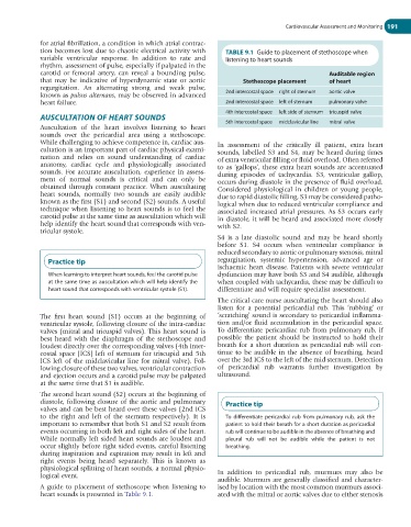
tion becomes lost due to chaotic electrical activity with TABLE 9.1 Guide to placement of stethoscope when
variable ventricular response. In addition to rate and listening to heart sounds
rhythm, assessment of pulse, especially if palpated in the
carotid or femoral artery, can reveal a bounding pulse, Auditable region
that may be indicative of hyperdynamic state or aortic Stethescope placement of heart
regurgitation. An alternating strong and weak pulse,
known as pulsus alternans, may be observed in advanced 2nd intercostal space right of sternum aortic valve
heart failure. 2nd intercostal space left of sternum pulmonary valve
4th intercostal space left side of sternum tricuspid valve
AUSCULTATION OF HEART SOUNDS
5th intercostal space midclavicular line mitral valve
Auscultation of the heart involves listening to heart
sounds over the pericardial area using a stethoscope.
While challenging to achieve competence in, cardiac aus- In assessment of the critically ill patient, extra heart
cultation is an important part of cardiac physical exami- sounds, labelled S3 and S4, may be heard during times
nation and relies on sound understanding of cardiac of extra ventricular filling or fluid overload. Often referred
anatomy, cardiac cycle and physiologically associated to as ‘gallops’, these extra heart sounds are accentuated
sounds. For accurate auscultation, experience in assess- during episodes of tachycardia. S3, ventricular gallop,
ment of normal sounds is critical and can only be occurs during diastole in the presence of fluid overload.
obtained through constant practice. When auscultating Considered physiological in children or young people,
heart sounds, normally two sounds are easily audible due to rapid diastolic filling, S3 may be considered patho-
known as the first (S1) and second (S2) sounds. A useful logical when due to reduced ventricular compliance and
technique when listening to heart sounds is to feel the associated increased atrial pressures. As S3 occurs early
carotid pulse at the same time as auscultation which will in diastole, it will be heard and associated more closely
help identify the heart sound that corresponds with ven- with S2.
tricular systole.
S4 is a late diastolic sound and may be heard shortly
before S1. S4 occurs when ventricular compliance is
reduced secondary to aortic or pulmonary stenosis, mitral
Practice tip regurgitation, systemic hypertension, advanced age or
ischaemic heart disease. Patients with severe ventricular
When learning to interpret heart sounds, feel the carotid pulse dysfunction may have both S3 and S4 audible, although
at the same time as auscultation which will help identify the when coupled with tachycardia, these may be difficult to
heart sound that corresponds with ventricular systole (S1). differentiate and will require specialist assessment.
The critical care nurse auscultating the heart should also
listen for a potential pericardial rub. This ‘rubbing’ or
The first heart sound (S1) occurs at the beginning of ‘scratching’ sound is secondary to pericardial inflamma-
ventricular systole, following closure of the intra-cardiac tion and/or fluid accumulation in the pericardial space.
valves (mitral and tricuspid valves). This heart sound is To differentiate pericardiac rub from pulmonary rub, if
best heard with the diaphragm of the stethoscope and possible the patient should be instructed to hold their
loudest directly over the corresponding valves (4th inter- breath for a short duration as pericardial rub will con-
costal space [ICS] left of sternum for triscupid and 5th tinue to be audible in the absence of breathing, heard
ICS left of the midclavicular line for mitral valve). Fol- over the 3rd ICS to the left of the mid sternum. Detection
lowing closure of these two valves, ventricular contraction of pericardial rub warrants further investigation by
and ejection occurs and a carotid pulse may be palpated ultrasound.
at the same time that S1 is audible.
The second heart sound (S2) occurs at the beginning of
diastole, following closure of the aortic and pulmonary Practice tip
valves and can be best heard over these valves (2nd ICS
to the right and left of the sternum respectively). It is To differentiate pericardial rub from pulmonary rub, ask the
important to remember that both S1 and S2 result from patient to hold their breath for a short duration as pericardial
events occurring in both left and right sides of the heart. rub will continue to be audible in the absence of breathing and
While normally left sided heart sounds are loudest and pleural rub will not be audible while the patient is not
occur slightly before right sided events, careful listening breathing.
during inspiration and expiration may result in left and
right events being heard separately. This is known as
physiological splitting of heart sounds, a normal physio-
logical event. In addition to pericardial rub, murmurs may also be
audible. Murmurs are generally classified and character-
A guide to placement of stethoscope when listening to ised by location with the most common murmurs associ-
heart sounds is presented in Table 9.1. ated with the mitral or aortic valves due to either stenosis