
Page 212 - ACCCN's Critical Care Nursing
P. 212
Cardiovascular Assessment and Monitoring 189
depressing these pacemaker cells. Cardiac innervation Capillary Capillary
includes sympathetic fibres from branches of T1–T5, and Endothelial cells network
10
parasympathetic input via the vagus nerve. The heart
rate at any moment is a product of the respective inputs
of sympathetic stimuli (which accelerate) and parasym-
pathetic stimuli (which depress) on heart rate. Rises in
heart rate can thus be achieved by an increase in sympa-
thetic tone or by a reduction in parasympathetic tone
(vagal inhibition). Conversely, slowing of the heart rate
can be achieved by decreasing sympathetic or increasing
parasympathetic activity. 4
Hormonal, biochemical and pharmacological inputs also
exert heart rate influences by their effect on autonomic Lumen
neural receptors or directly on pacemaker cells. In Valve
mimicking the effects of direct nervous inputs, these Tunica intima:
influences may be described as sympathomimetic or Endothelium
parasympathomimetic. Sympathomimetic stimulation Subendothelial layer
(e.g. through the use of isoprenaline) achieves the same Internal elastic lamina
cardiac endpoints as direct sympathetic activity, increas- Artery Vein
ing the heart rate, while sympathetic antagonism (e.g. Tunica media
beta-blockade therapy) slows the heart through receptor Tunica adventitia
inhibition. By contrast, parasympathomimetic agonist 3
activity slows the heart rate, while parasympathetic anta- FIGURE 9.11 The structure of arteries, veins and capillaries.
gonism (e.g. via administration of atropine sulphate)
raises the heart rate by causing parasympathetic receptor
blockade. 4
veins are numerous and have thinner, less muscular walls,
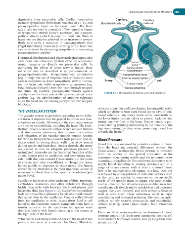
THE VASCULAR SYSTEM which can dilate to store extra blood (up to 64% of total
The vascular system is specialised according to the differ- blood volume at any time). Some veins, particularly in
ent tissue it supplies, but the general functions and char- the lower limbs, contain valves to prevent backflow and
acteristics are similar. All vessels in the circulatory system ensure one-way flow to the heart. Venous return is pro-
are lined by endothelium, including the heart. The endo- moted during standing and moving by the muscles of the
thelium creates a smooth surface, which reduces friction legs compressing the deep veins, promoting blood flow
and also secretes substances that promote contraction towards the heart. 1,4
and relaxation of the vascular smooth muscle. Arteries
function to transport blood under high pressure and are Blood Pressure
characterised by strong elastic walls that allow stretch Blood flow is maintained by pulsatile ejection of blood
during systole and high flow. During diastole, the artery from the heart and pressure differences between the
walls recoil so that an adequate perfusion pressure is blood vessels. Traditionally, blood pressure is measured
maintained. Arterioles are the final small branches of the from the arteries in the general circulation at the
arterial system prior to capillaries, and have strong mus- maximum value during systole and the minimum value
cular walls that can contract (vasoconstrict) to the point occurring during diastole. The cardiovascular system must
of closure and relax (vasodilate) to change the artery supply blood according to varying demands and in a
lumen rapidly in response to tissue needs. The lumen range of circumstances, with at least a minimal blood
created by the arterioles is the most important source of flow to be maintained to all organs. At a local level this
resistance to blood flow in the systemic circulation (just is achieved by autoregulation of individual arteries, such
under 50%).
as the coronary arteries, in response to the metabolic
Capillaries function to allow exchange of fluid, nutrients, needs of the specific tissue or organ. The exact mecha-
electrolytes, hormones and other substances through nism is unknown, but it has been proposed that increased
highly permeable walls between the blood plasma and vascular muscle stretch and/or metabolites and decreased
interstitial fluid (see Figure 9.11). Just before the capillary oxygen levels are detected and cells release substances
beds are precapillary sphincters, bands of smooth muscle such as adenosine. These substances result in rapid
4
that adjust flow in the capillaries. Venules collect blood vasodilation and increased perfusion. The vascular endo-
from the capillaries to veins. Excess tissue fluid is col- thelium actively secretes prostacyclin and endothelial-
lected by the lymphatic system. Lymphatic veins have a derived relaxing factor (nitric oxide), both vasoactive
similar structure to the cardiovascular system veins agents.
described below, with lymph returning to this system at
the right side of the heart. There are three main regulatory mechanisms of blood
pressure control: (a) short-term autonomic control; (b)
Veins collect and transport blood back to the heart at low medium-term hormonal control; and (c) long-term renal
pressure and serve as a reservoir for blood. Therefore, system control.