Page 449 - ACCCN's Critical Care Nursing
P. 449

426  P R I N C I P L E S   A N D   P R A C T I C E   O F   C R I T I C A L   C A R E


                            Extension of choroid                                                  Cranium
                            plexus into lateral ventricle                                         Dura mater
             Choroid plexus                         Arachnoid                                     (endosteal
             of third ventricle                     granulations                                  layer)

                                                                                                  Fluid
                                                                                                  movement
                                                                                                  Superior
                                                                                                  sagittal sinus
                                                                                                  Arachnoid
                                                                                                  granulation
                                                                                                  Dura mater
                                                                                                  (meningeal
                                                                                                  layer)
                                                                   Cerebral  Subarachnoid Arachnoid  Subdural
                                                                     cortex        space              space
                                                                          Pia
                                                                          mater
                                                              Superior
                                                              sagittal
                Mesencephalic                                 sinus
                    aqueduct
                   Lateral aperture                               B
                   Choroid plexus of
                   fourth ventricle              Spinal
                     Median aperture             cord
                          Arachnoid             Central canal
                   Subarachnoid space

                          Dura mater


                                                  Filum
                                                  terminale


                   A

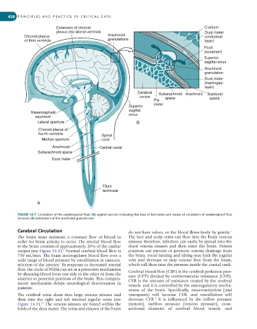
         FIGURE 16.7  Circulation of the cerebrospinal fluid: (A) sagittal section indicating the sites of formation and routes of circulation of cerebrospinal fluid
         (arrows); (B) orientation of the arachnoid granulations.   1


         Cerebral Circulation                                 do not have valves, so the blood flows freely by gravity.
                                                                                                               1
         The  brain  must  maintain  a  constant  flow  of  blood  in   The face and scalp veins can flow into the brain venous
         order for brain activity to occur. The arterial blood flow   sinuses; therefore, infection can easily be spread into the
         to the brain consists of approximately 20% of the cardiac   dural  venous  sinuses  and  then  enter  the  brain.  Patient
         output (see Figure 16.8).  Normal cerebral blood flow is   position can prevent or promote venous drainage from
                               5
         750 mL/min. The brain autoregulates blood flow over a   the brain. Head turning and tilting may kink the jugular
         wide range of blood pressure by vasodilation or vasocon-  vein  and  decrease  or  stop  venous  flow  from  the  brain,
         striction of the arteries.  In response to decreased arterial   which will then raise the pressure inside the cranial vault.
                             1
         flow, the circle of Willis can act as a protective mechanism   Cerebral blood flow (CBF) is the cerebral perfusion pres-
         by shunting blood from one side to the other or from the   sure (CPP) divided by cerebrovascular resistance (CVR).
         anterior to posterior portions of the brain. This compen-  CVR is the amount of resistance created by the cerebral
         satory  mechanism  delays  neurological  deterioration  in   vessels, and it is controlled by the autoregulatory mecha-
         patients.
                                                              nisms  of  the  brain.  Specifically,  vasoconstriction  (and
         The  cerebral  veins  drain  into  large  venous  sinuses  and   vasospasm)  will  increase  CVR,  and  vasodilation  will
                                                                           1
         then  into  the  right  and  left  internal  jugular  veins  (see   decrease  CVR.   It  is  influenced  by  the  inflow  pressure
                     23
         Figure 16.9).  The venous sinuses are found within the   (systole),  outflow  pressure  (venous  pressure),  cross-
         folds of the dura mater. The veins and sinuses of the brain   sectional  diameter  of  cerebral  blood  vessels,  and
   444   445   446   447   448   449   450   451   452   453   454