
Page 448 - ACCCN's Critical Care Nursing
P. 448
Neurological Assessment and Monitoring 425
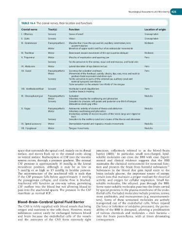
TABLE 16.4 The cranial nerves, their location and functions
Cranial nerve Tract(s) Function Location of origin
I. Olfactory Sensory Sense of smell Diencephalon
II. Optic Sensory Vision Diencephalon
III. Oculomotor Parasympathetic Muscles that move the eye and lid, pupillary constriction, lens Midbrain
accommodation
Motor Elevation of upper eyelid and four of six extraocular movements
IV. Trochlear Motor Downward, inward movement of the eye (superior oblique) Midbrain
V. Trigeminal Motor Muscles of mastication and opening jaw Pons
Sensory Tactile sensation to the cornea, nasal and oral mucosa, and facial skin
VI. Abducens Motor Lateral deviation of eye (lateral rectus) Pons
VII. Facial Parasympathetic Secretory for salivation and tears Pons
Motor Movement of the forehead, eyelids, cheeks, lips, ears, nose and neck to
produce facial expression and close eyes
Sensory Tactile sensation to parts of the external ear, auditory canal and
external tympanic membrane
Taste sensation to the anterior two-thirds of the tongue
VIII. Vestibulocochlear Sensory Vestibular branch: Equilibrium Pons
Cochlear branch: Hearing
IX. Glossopharyngeal Parasympathetic Salivation Medulla
Motor Voluntary muscles for swallowing and phonation
Sensory Sensation to pharynx, soft palate and posterior one-third of tongue
Stimulation elicits gag reflex
X. Vagus Parasympathetic Autonomic activity of viscera of thorax and abdomen Medulla
Motor Voluntary swallowing and phonation
Involuntary activity of visceral muscles of the heart, lungs and digestive
tract
Sensory Sensation to the auditory canal and viscera of the thorax and abdomen
XI. Spinal accessory Motor Sternocleidomastoid and trapezius muscle movements Medulla
XII. Hyoglossal Motor Tongue movements Medulla
space that surrounds the spinal cord, mainly on its dorsal junctions, collectively referred to as the blood–brain
1
surface, and moves back up to the cranial cavity along barrier (BBB). In particular, small non-charged, lipid-
its ventral surface. Reabsorption of CSF into the vascular soluble molecules can cross the BBB with ease. Experi-
system occurs, through a pressure gradient. The normal mental and clinical evidence suggests that the BBB
CSF pressure is approximately 10 mmHg in the lateral maintains the chemical environment for neuronal func-
22
recumbent position, although it may be as low as tion and protects the brain from harmful substances.
5 mmHg or as high as 15 mmHg in healthy persons. Substances in the blood that gain rapid entry to the
The microstructure of the arachnoid villi is such that brain include glucose, the important source of energy,
if the CSF pressure falls below approximately 3 mmHg certain ions that maintain a proper medium for electrical
the passageways collapse, and reverse flow is blocked. activity, and oxygen for cellular respiration. Small fat-
Arachnoid villi function as one-way valves, permitting soluble molecules, like ethanol, pass through the BBB.
CSF outflow into the blood but not allowing blood to Some water-soluble molecules pass into the brain carried
pass into the arachnoid spaces. The pressure in the CSF by special proteins in the plasma membrane of the endo-
manifests as normal ICP. thelial cells. Excluded molecules include proteins, toxins,
most antibiotics, and monoamines (e.g. neurotransmit-
ters). Some of these unwanted molecules are actively
Blood–Brain–Cerebral Spinal Fluid Barrier transported out of the endothelial cells. When injured
The CNS is richly supplied with blood vessels that bring (by force or infection or oxidative processes), the perme-
oxygen and nutrients to the cells there. However, many ability of the BBB is disrupted, allowing a proliferation
substances cannot easily be exchanged between blood of various chemicals and molecules – even bacteria –
and brain because the endothelial cells of the vessels into the brain parenchyma, with at times devastating
and the astrocytes of the CNS form extremely tight consequences.