
Page 447 - ACCCN's Critical Care Nursing
P. 447
424 P R I N C I P L E S A N D P R A C T I C E O F C R I T I C A L C A R E
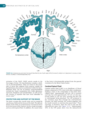
Knee Hip Trunk Shoulder Arm
Leg
Elbow Wrist Hand Fingers
Head
Hip
Neck
Trunk
Arm
Elbow
Forearm
Thumb
Hand
Fingers
Neck
Thumb
Eye Brow
Eye
Nose
Face
Face
Lips Midline Toes Lips
Genitals
Teeth Jaw
Gums
Jaw Tongue
Tongue Pharynx
Pharynx Larynx
Abdomen
Somatosensory cortex Motor cortex
Left Right
FIGURE 16.6 Somatosensory and motor homunculi. Note that the size of each region of the homunculi is related to its importance in sensory or motor
15
function, resulting in a distorted-appearing map.
activation of the DMTF. DMTF activity results in the of the brain is biochemically isolated from the general
four stages of sleep. The hypothalamus contains a pleth- circulation by the blood–brain barrier.
ora of neurotransmitters. These are found in the termi-
nals of axons that originate from neurons outside the Cerebral Spinal Fluid
hypothalamus, but most are synthesised within the hypo- Cerebral spinal fluid (CSF) is an ultrafiltrate of blood
thalamus itself. The list of putative neurotransmitters plasma composed of 99% water with other constituents,
includes the ‘classic’ transmitters ACh, GABA, glutamate, making it close to the composition of the brain extra-
serotonin, dopamine and noradrenaline, as well as liter- cellular fluid. Approximately 500 mL CSF is secreted
1
ally dozens of peptides that have been identified in each day, but only approximately 150 mL is in the ven-
recent years. 20 tricular system at any one time, meaning that the CSF
is continuously being absorbed. The CSF produced in
PROTECTION AND SUPPORT OF THE BRAIN the ventricles must flow through the interventricular
The brain occupies the cranial cavity and is covered by foramen, the third ventricle, the cerebral aqueduct and
21
membranes, fluid and the bones of the skull. The delicate the fourth ventricle to exit from the neural tube. Three
tissues of the brain are protected from mechanical forces openings, or foramina, allow the CSF to pass into the
1
by (a) the bones of the cranium, (b) the cranial meninges, subarachnoid space (see Figure 16.7). Approximately
and (c) cerebrospinal fluid. In addition, the neural tissue 30% of the CSF passes down into the subarachnoid