
Page 517 - ACCCN's Critical Care Nursing
P. 517
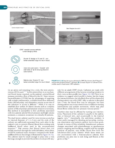
494  P R I N C I P L E S   A N D   P R A C T I C E   O F   C R I T I C A L   C A R E


           A
                      CRRT vascular access catheter
                         lumen design profiles


                          Double ‘D’ design or ‘D’ and ‘O’ : one
                          lumen extended longer for return blood


                          Inner and outer lumen : ‘Coaxial’  with
                          side holes at  tip for drawing blood int
                          outer lumeno


                          Side by side : Double ‘O’ :one  FIGURE 18.15  (A) Vascular access catheters for CRRT. Dual-lumen, Bard® Niagara™
                          lumen extended longer for return blood  and Gambro Dolphin Protect™ catheters; (B) Concept diagram of catheter lumen
           B                                            profiles used for dual lumen CRRT catheters.


         via an artery and returning it to a vein, the term arterio-  min for an adult CRRT circuit. Catheters are made with
         venous (AV) is used. 75,76  In this system there is no mechan-  different arrangement of the lumens revealing variation in
         ical blood pump required, as the patient’s arterial blood   their cross section profile (see Figure 18.15b) There is no
         pressure provides a flow of blood in the EC. Veno-venous   evidence to suggest which profile is better, but the larger
         haemofiltration (CVVH) has the advantages of requiring   the internal diameter, the less likely flow will be obstructed
         only a single venipuncture, a reliable blood flow delivered   during patient care with CRRT. After a catheter is threaded
         from a blood pump, and alternative venous access sites if   into a vein, the blood flow may be adequate, but later
                                        77
         site  infection  or  access  is  difficult.   While  it  is  easy  to   during patient care it may obstruct due to different nursing
         establish flow within AV-driven circuits and no complex   interventions  and  patient  movement,  which  may  alter
         system of blood pumps and pressure sensors is necessary,   blood flows within the low pressure venous system. 70
         this  method  is  susceptible  to  flow  problems  associated
         with low patient arterial blood pressure and high venous   Insertion sites may be affected by nursing care interven-
         pressures, a common occurrence in critically ill patients.  tions. Placement of the catheter is usually in the subcla-
                                                              vian  or  femoral  vein,  and  occasionally  in  the  internal
                                                                          78
         The dual-lumen catheter used for veno-venous access has   jugular  vein.   Anecdotally,  the  subclavian  position  is
         an internal diameter of 1.5–3 mm and the ends of the   more easily managed for dressing and securing, continu-
         catheter are sufficiently separated from each other in the   ous observation and patient comfort, but is more prob-
         patient’s vein to prevent filtered blood from mixing with   lematic in terms of flow reliability. Intrathoracic pressure
         unfiltered  blood  when  used  in  the  recommended   changes  associated  with  physiotherapy  or  spontaneous
                  77
         sequence.   This  ensures  that  filtered  blood  does  not   patient coughing and breathing, coupled with the upright
         simply pass back through the artificial kidney, where there   position  of  patients,  may  hinder  blood  flow  from  the
         would be minimal waste clearance compared with ‘fresh’   subclavian-sited  access  catheter.  While  these  issues  are
         unfiltered blood; this design is illustrated in Figure 18.15a.   not  encountered  with  a  femoral-placed  catheter,  flow
         The catheter must be small enough to place into a vein   problems can arise due to side lying and flexion at the
         but large enough to provide blood flow of at least 200 mL/  groin or hip. 42