


Page 52 - Concise Pathology for Exam Preparation ( PDFDrive )
P. 52
2  Acute and Chronic Inflammation  37

             Steps in Phagocytosis (Fig. 2.4)

               1.  Recognition and attachment
                 (a)  Typically,  phagocytosis  is  initiated  by  recognition  of  the  microorganisms  and
                   particles by receptors expressed on the leukocyte surface.
                 (b)  Mannose receptors and scavenger receptors are two important receptors that
                   function to bind and ingest microbes. Mannose/fucose residues are typically a part
                   of the microbial cell wall; whereas, mammalian cells instead contain sialic acid and
                   N-acetylgalactosamine  residues.  Mannose  receptors,  therefore,  recognize  only
                   the microbe and not the host cell. Macrophage scavenger receptors bind a lot of
                   microbes.
                 (c)  The efficiency of phagocytosis is greatly enhanced by opsonization of bacteria (or
                   foreign material).
                  (d)  The process of coating of a particle, such as a microbe, to target it for phagocytosis
                   is called opsonization and the substances that do this are called opsonins. Phago-
                   cytes express high-affinity receptors for opsonins.
                 (e)  Major opsonins are ‘IgG antibodies’, ‘C3b breakdown products of complement’
                   and plasma carbohydrate-binding lectins called ‘collectins’, which bind to the
                   microbial cell wall sugar groups.
                 (f)  Leukocytes express receptors for opsonins that facilitate phagocytosis of the coated
                   microbes, eg, Fc receptor for IgG (FcgRI), complement receptors 1 and 3 (CR1
                   and 3) for complement fragments and C1q for the collectins.
               2.  Engulfment
                 (a)  Bacteria  are  engulfed  by  pseudopodia  (extensions  of  cytoplasm)  and  trapped
                   within phagosomes forming a phagocytic vacuole.
                 (b)  The limiting membrane of the phagocytic vacuole fuses with the limiting mem-
                   brane of the lysosomal granule, resulting in discharge of the contents of the granule
                   into the phagolysosome.
               3.  Killing and degradation
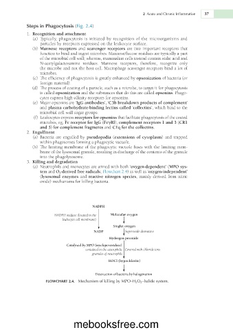
                 (a)  Neutrophils and monocytes are armed with both ‘oxygen-dependent’ (MPO sys-
                   tem and O 2 -derived free radicals; Flowchart 2.4) as well as ‘oxygen-independent’
                   (lysosomal enzymes and reactive nitrogen species, mainly derived from nitric
                   oxide) mechanisms for killing bacteria.


                                          NADPH

                         NADPH oxidase (located in the    Molecular oxygen
                             leukocyte cell membrane)
                                                    Singlet oxygen
                                           NADP          Superoxide dismutase
                                                  Hydrogen peroxide
                               Catalysed by MPO (myeloperoxidase)
                                       contained in the azurophilic  Covered with chloride ions
                                          granules of neutrophils
                                                  HOCl (hypochlorite)


                                            Destruction of bacteria by halogenation
                      FLOWCHART 2.4.  Mechanism of killing by MPO–H 2 O 2 –halide system.


                                  mebooksfree.com

