
Page 1045 - Hematology_ Basic Principles and Practice ( PDFDrive )
P. 1045
928 Part VII Hematologic Malignancies
B
A C
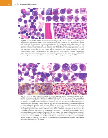
Fig. 59.4 ACUTE MYELOID LEUKEMIA WITH t(8;21)(q22;q22), (RUNX1-RUNX1T1). (A) Low-power,
Wright-stained bone marrow aspirate smear showing increased blasts associated with differentiating myeloid
cells. (B) Details illustrating some of the features associated with this leukemia. They include blasts with long
thin Auer rods (top left), immature cells with abnormal eosinophilic globules (top and bottom, second from left),
abnormal salmon-colored granulation in the maturing cells, sometimes associated with a basophilic periphery
(top and bottom, fourth from left), and slightly abnormal features in the mature neutrophils (far right).
Pseudo–Chediak-Higashi granules were not seen in this case. (C) Biopsy specimen illustrates the significant
degree of maturation that can sometimes be seen. In fact, in some cases the blast count can be less than 20%,
but the diagnosis of acute myeloid leukemia is still made with the cytogenetic finding of t(8;21).
B B
A C C DD
E E F
Fig. 59.5 ACUTE MYELOID LEUKEMIA WITH ABNORMAL BONE MARROW EOSINOPHILS
AND INV(16)(p13.1;q22) OR t(16;16)(p13.1;q22), (CBFB-MYH11). (A) Low-power, Wright-stained bone
marrow aspirate showing blasts, monocytic cells, granulocytic cells, and abnormal eosinophils. (B) Features of
the abnormal eosinophils in three abnormal eosinophils (left three cells). Note the abnormal basophilic granules
in the eosinophilic myelocytes. These granules are large, tend to cluster or coalesce, and are interspersed among
the large eosinophilic granules, which are more difficult to see. As the eosinophils mature, the abnormal
basophilic granules are less prominent and sometimes disappear (far right). A common misconception is that
the basophilic granules are the granules of basophils and that the abnormal eosinophils are “hybrid cells.” (C)
A basophil in the same case (top cell) for comparison. (D) Cells from a case of reactive eosinophilia, also for
comparison. Immature eosinophils (cell to the right) do have primary blue granules. However, they are usually
less prominent and less atypical than the basophilic granules of the abnormal eosinophils. It is notable that
the monocytes in cases of AML with inv(16) or t(16;16) are sometimes α-naphthyl acetate esterase (ANAE)
reaction negative. (E) Negative ANAE reaction. (F) Abnormal eosinophils cannot be recognized in hematoxylin
and eosin-stained sections of the biopsy.