
Page 1046 - Hematology_ Basic Principles and Practice ( PDFDrive )
P. 1046
Chapter 59 Clinical Manifestations and Treatment of Acute Myeloid Leukemia 929
A A B B C C GG
D D E E F F
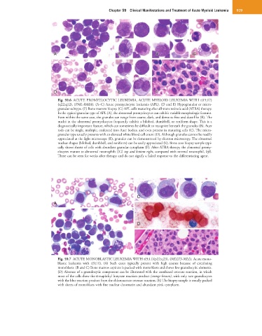
Fig. 59.6 ACUTE PROMYELOCYTIC LEUKEMIA, ACUTE MYELOID LEUKEMIA WITH t(15;17)
(q22;q12), (PML-RARA). (A–C) Acute promyelocytic leukemia (APL). (D and E) Hypogranular or micro-
granular subtype. (F) Bone marrow biopsy. (G) APL cells maturing after all-trans retinoic acid (ATRA) therapy.
In the typical granular type of APL (A), the abnormal promyelocytes can exhibit variable morphologic features.
Even within the same case, the granules can range from coarse, dark, and dense to fine and dust-like (B). The
nuclei in the abnormal promyelocytes frequently exhibit a bilobed, dumbbell, or reniform shape. This is a
diagnostically important feature, which can sometimes be difficult to recognize beneath the granules (B). Auer
rods can be single, multiple, coalesced into Auer bodies, and even present in maturing cells (C). The micro-
granular type usually presents with an elevated white blood cell count (D). Although granules cannot be readily
appreciated at the light microscope (E), granules can be demonstrated by electron microscopy. The abnormal
nuclear shapes (bilobed, dumbbell, and reniform) can be easily appreciated (E). Bone core biopsy sample typi-
cally shows sheets of cells with abundant granular cytoplasm (F). After ATRA therapy the abnormal promy-
elocytes mature to abnormal neutrophils ([G] top and bottom right, compared with normal neutrophil, left).
These can be seen for weeks after therapy and do not signify a failed response to the differentiating agent.
C
A
D
B E
Fig. 59.7 ACUTE MONOBLASTIC LEUKEMIA WITH t(9;11)(p22;q23), (MLLT3-MLL). Acute mono-
blastic leukemia with t(9;11). (A) Such cases typically present with high counts because of circulating
monoblasts. (B and C) Bone marrow aspirate is packed with monoblasts and shows few granulocytic elements.
(D) Absence of a granulocytic component can be illustrated with the combined esterase reaction, in which
most of the cells show the α-naphthyl butyrate reaction product (orange-brown), with only rare granulocytes
with the blue reaction product from the chloroacetate esterase reaction. (E) The biopsy sample is usually packed
with sheets of monoblasts with fine nuclear chromatin and abundant pink cytoplasm.