
Page 1348 - Hematology_ Basic Principles and Practice ( PDFDrive )
P. 1348
1194 Part VII Hematologic Malignancies
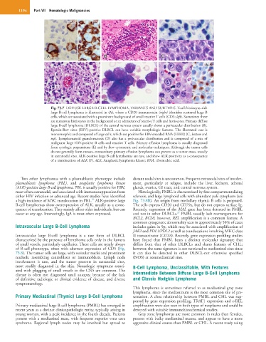
CD20 CD3
A A B B CC
ALK
D D E E F F
Fig. 73.7 DIFFUSE LARGE B-CELL LYMPHOMA, VARIANTS AND SUBTYPES. T-cell/histiocyte–rich
large B-cell lymphoma is illustrated in (A), where a CD20 immunostain (right) identifies scattered large B
cells, which are associated with a prominent background of small reactive T cells (CD3; left). Sometimes there
are numerous histiocytes in the background or an admixture of reactive T cells and histiocytes. Primary diffuse
large B-cell lymphoma (DLBCL) of the central nervous system usually shows a perivascular distribution (B).
Epstein-Barr virus (EBV)–positive DLBCL can have variable morphologic features. The illustrated case is
monomorphic and composed of large cells, which are positive for EBV-encoded RNA (EBER) (C, bottom and
top). Lymphomatoid granulomatosis (D) also has a perivascular distribution and is composed of a mix of
malignant large EBV-positive B cells and reactive T cells. Primary effusion lymphoma is usually diagnosed
from cytologic preparations (E) and by flow cytometric and molecular techniques. Although the tumor cells
do not generally form masses, extracavitary primary effusion lymphoma can present as a tumor mass, usually
in extranodal sites. ALK-positive large B-cell lymphomas are rare, and show ALK positivity as a consequence
of a translocation of ALK (F). ALK, Anaplastic lymphoma kinase; RNA, ribonucleic acid.
Two other lymphomas with a plasmablastic phenotype include distant nodal sites is uncommon. Frequent extranodal sites of involve-
plasmablastic lymphoma (PBL), and anaplastic lymphoma kinase ment, particularly at relapse, include the liver, kidneys, adrenal
(ALK)-positive large B-cell lymphoma. PBL is usually positive for EBV, glands, ovaries, GI tract, and central nervous system.
most often extranodal, and associated with immunosuppression from Histologically, PMBL is characterized by fine compartmentalizing
either HIV infection or advanced age. Recent studies have identified sclerosis, and large lymphoid cells with abundant pale cytoplasm (see
26
a high incidence of MYC translocation in PBL. ALK-positive large Fig. 73.8B). An origin from medullary thymic B cells is proposed.
B-cell lymphomas show overexpression of ALK, usually as a conse- The cells express CD20 and CD79a, but do not express surface Ig.
quence of translocation. They mainly affect older individuals, but can Recently, expression of the MAL gene has been detected in PMBL
27
occur at any age. Interestingly, IgA is most often expressed. and not in other DLBCLs. PMBL usually lack rearrangement for
BCL2, BCL6; however, REL amplification is a common feature. A
common cytogenetic abnormality seen in approximately 50% of cases
Intravascular Large B-Cell Lymphoma includes gains in 9p, which may be associated with amplification of
JAK2 and PDL1/PDL2 as well as translocations involving MHC class
Intravascular large B-cell lymphoma is a rare form of DLBCL II transactivator (CIITA). Recently, gene expression profiling studies
characterized by the presence of lymphoma cells only in the lumens have found that PMBL bears a distinct molecular signature that
of small vessels, particularly capillaries. These cells are nearly always differs from that of other DLBCLs and shares features of CHL;
of B-cell phenotype, often with aberrant expression of CD5 (Fig. however, the same signature is not restricted to mediastinal sites since
73.8). The tumor cells are large, with vesicular nuclei and prominent it can also be detected in other DLBCL-not otherwise specified
nucleoli, resembling centroblasts or immunoblasts. Lymph node (NOS) at nonmediastinal sites.
involvement is rare, and the tumor presents in extranodal sites,
most readily diagnosed in the skin. Neurologic symptoms associ- B-Cell Lymphoma, Unclassifiable, With Features
ated with plugging of small vessels in the CNS are common. The
disease is often not diagnosed until autopsy, because of the lack Intermediate Between Diffuse Large B-Cell Lymphoma
of definitive radiologic or clinical evidence of disease, and diverse and Classic Hodgkin Lymphoma
symptomatology.
This lymphoma is sometimes referred to as mediastinal gray zone
lymphoma, since the mediastinum is the most common site of pre-
Primary Mediastinal (Thymic) Large B-Cell Lymphoma sentation. A close relationship between PMBL and CHL was sup-
ported by gene expression profiling. TRAF1 expression and c-REL
Primary mediastinal large B-cell lymphoma (PMBL) has emerged in amplification were also seen in both types of neoplasms and could be
recent years as a distinct clinicopathologic entity, typically arising in detected with suitable immunohistochemical studies.
young women, with a peak incidence in the fourth decade. Patients Gray zone lymphomas are more common in males than females,
present with a mediastinal mass, with frequent superior vena cava present with bulky mediastinal masses, and appear to have a more
syndrome. Regional lymph nodes may be involved but spread to aggressive clinical course than PMBL or CHL. A recent study using