
Page 1353 - Hematology_ Basic Principles and Practice ( PDFDrive )
P. 1353
Chapter 73 The Pathologic Basis for the Classification of Non-Hodgkin and Hodgkin Lymphomas 1199
A A B C
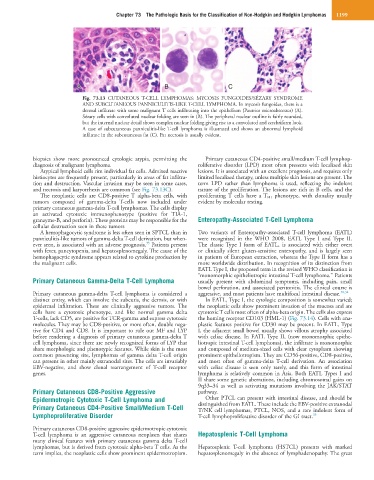
Fig. 73.13 CUTANEOUS T-CELL LYMPHOMAS: MYCOSIS FUNGOIDES/SÉZARY SYNDROME
AND SUBCUTANEOUS PANNICULITIS-LIKE T-CELL LYMPHOMA. In mycosis fungoides, there is a
dermal infiltrate with some malignant T cells infiltrating into the epithelium (Pautrier microabscesses) (A).
Sézary cells with convoluted nuclear folding are seen in (B). The peripheral nuclear outline is fairly rounded,
but the internal nuclear detail shows complex nuclear folding giving rise to a convoluted and cerebriform look.
A case of subcutaneous panniculitis-like T-cell lymphoma is illustrated and shows an abnormal lymphoid
infiltrate in the subcutaneous fat (C). Fat necrosis is usually evident.
biopsies show more pronounced cytologic atypia, permitting the Primary cutaneous CD4-positive small/medium T-cell lymphop-
diagnosis of malignant lymphoma. roliferative disorder (LPD) most often presents with localized skin
Atypical lymphoid cells rim individual fat cells. Admixed reactive lesions. It is associated with an excellent prognosis, and requires only
histiocytes are frequently present, particularly in areas of fat infiltra- limited localized therapy, unless multiple skin lesions are present. The
tion and destruction. Vascular invasion may be seen in some cases, term LPD rather than lymphoma is used, reflecting the indolent
and necrosis and karyorrhexis are common (see Fig. 73.13C). nature of the proliferation. The lesions are rich in B cells, and the
The neoplastic cells are CD8-positive T alpha-beta cells, with proliferating T cells have a T FH phenotype, with clonality usually
tumors composed of gamma-delta T-cells now included under evident by molecular testing.
primary cutaneous gamma-delta T-cell lymphomas. The cells display
an activated cytotoxic immunophenotype (positive for TIA-1,
granzyme-B, and perforin). These proteins may be responsible for the Enteropathy-Associated T-Cell Lymphoma
cellular destruction seen in these tumors
A hemophagocytic syndrome is less often seen in SPTCL than in Two variants of Enteropathy-associated T-cell lymphoma (EATL)
panniculitis-like tumors of gamma-delta T-cell derivation, but when- were recognized in the WHO 2008, EATL Type I and Type II.
33
ever seen, is associated with an adverse prognosis. Patients present The classic Type I form of EATL, is associated with either overt
with fever, pancytopenia, and hepatosplenomegaly. The cause of the or clinically silent gluten-sensitive enteropathy, and is largely seen
hemophagocytic syndrome appears related to cytokine production by in patients of European extraction, whereas the Type II form has a
the malignant cells. more worldwide distribution. In recognition of its distinction from
EATL Type I, the proposed term in the revised WHO classification is
“monomorphic epitheliotropic intestinal T-cell lymphoma.” Patients
Primary Cutaneous Gamma-Delta T-Cell Lymphoma usually present with abdominal symptoms, including pain, small
bowel perforation, and associated peritonitis. The clinical course is
Primary cutaneous gamma-delta T-cell lymphoma is considered a aggressive, and most patients have multifocal intestinal disease. 30,34
distinct entity, which can involve the subcutis, the dermis, or with In EATL, Type I, the cytologic composition is somewhat varied;
epidermal infiltration. These are clinically aggressive tumors. The the neoplastic cells show prominent invasion of the mucosa and are
cells have a cytotoxic phenotype, and like normal gamma delta cytotoxic T cells most often of alpha-beta origin. The cells also express
T-cells, lack CD5, are positive for TCR-gamma and express cytotoxic the homing receptor CD103 (HML-1) (Fig. 73.14). Cells with ana-
molecules. They may be CD8-positive, or more often, double nega- plastic features positive for CD30 may be present. In EATL, Type
tive for CD4 and CD8. It is important to rule out MF and LYP I, the adjacent small bowel usually shows villous atrophy associated
before rendering a diagnosis of primary cutaneous gamma-delta T with celiac disease. In EATL Type II, (now monomorphic epithe-
cell lymphoma, since there are newly recognized forms of LYP that liotropic intestinal T-cell lymphoma), the infiltrate is monomorphic
share morphologic and phenotypic features. While skin is the most and composed of medium-sized cells with clear cytoplasm showing
common presenting site, lymphomas of gamma delta T-cell origin prominent epitheliotropism. They are CD56-positive, CD8-positive,
can present in other mainly extranodal sites. The cells are invariably and most often of gamma-delta T-cell derivation. An association
EBV-negative, and show clonal rearrangement of T-cell receptor with celiac disease is seen only rarely, and this form of intestinal
genes. lymphoma is relatively common in Asia. Both EATL Types I and
II share some genetic aberrations, including chromosomal gains on
9q33–34 as well as activating mutations involving the JAK/STAT
Primary Cutaneous CD8-Positive Aggressive pathway.
Epidermotropic Cytotoxic T-Cell Lymphoma and Other PTCL can present with intestinal disease, and should be
Primary Cutaneous CD4-Positive Small/Medium T-Cell distinguished from EATL. These include the EBV-positive extranodal
T/NK cell lymphomas, PTCL, NOS, and a rare indolent form of
Lymphoproliferative Disorder T-cell lymphoproliferative disorder of the GI tract. 35
Primary cutaneous CD8-positive aggressive epidermotropic cytotoxic
T-cell lymphoma is an aggressive cutaneous neoplasm that shares Hepatosplenic T-Cell Lymphoma
many clinical features with primary cutaneous gamma delta T-cell
lymphomas, but is derived from cytotoxic alpha-beta T cells. As the Hepatosplenic T-cell lymphoma (HSTCL) presents with marked
term implies, the neoplastic cells show prominent epidermotropism. hepatosplenomegaly in the absence of lymphadenopathy. The great