
Page 1351 - Hematology_ Basic Principles and Practice ( PDFDrive )
P. 1351
Chapter 73 The Pathologic Basis for the Classification of Non-Hodgkin and Hodgkin Lymphomas 1197
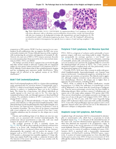
A A B C C
Fig. 73.11 PERIPHERAL T-CELL LYMPHOMAS. In angioimmunoblastic T-cell lymphoma, the lymph
node shows effacement with an arborizing vascular proliferation of postcapillary venules and clustered large
cells with clear cytoplasm in the background of plasma cells, immunoblasts, and small lymphocytes (A). The
peripheral blood in adult T-cell leukemia/lymphoma has classic “flower-cells” (B). Peripheral T-cell lymphoma,
not otherwise specified is heterogeneous, but typically there is a mixture of small and large neoplastic T cells
(C).
progression to EBV-positive DLBCL has been reported in rare cases. Peripheral T-Cell Lymphomas, Not Otherwise Specified
Atypical B-cell proliferations that are negative for EBV also occur,
presumably related to the T-helper follicular function of the neoplastic PTCL, NOS is a diagnosis of exclusion and is admittedly a hetero-
cells in promoting the activation and migration of bystander B cells. geneous category with most cases being nodal in origin. Therefore,
30
However, the exact role of EBV in AITL remains uncertain. Recently not unexpectedly the cytologic spectrum is very broad. (see
recurrent mutations have been reported in AITL; these include muta- Fig. 73.11C). An inflammatory background is frequent, consisting
tions in IDH2, TET2, and RHOA. of eosinophils, plasma cells, and histiocytes. If the epithelioid histio-
The biologic spectrum of AITL is greater than previously thought cytes are numerous and clustered, the neoplasm fulfils the criteria for
and other entities related to follicular T-helper cells are considered the lymphoepithelioid cell variant of PTCL. The T-zone variant is
within the same group. Indeed, peripheral T-cell lymphoma (PTCL)- composed of small to medium-sized cells that preferentially involve
NOS, follicular variant shares some of the underlying genetic changes the paracortical regions of the lymph node.
and has overlapping gene expression profile characteristics. These Clinically, PTCL, NOS most often presents in adults with gener-
changes will be reflected in the updated version of the WHO alized lymphadenopathy, hepatosplenomegaly, and frequent bone
classification. 30 marrow involvement. Constitutional symptoms, including fever and
night sweats, are common, as is pruritus. The clinical course is aggres-
sive, although complete remissions may be obtained with combina-
Adult T-Cell Leukemia/Lymphoma tion chemotherapy. However, the relapse rate is higher than in
aggressive B-cell lymphomas, including DLBCL.
Adult T-cell leukemia/lymphoma (ATL) is a distinct clinicopathologic PTCL, NOS, as defined in the WHO classification, remains
entity associated with the retrovirus human T-lymphotropic virus-1 heterogeneous. It is likely that individual clinicopathologic entities
(HTLV-1), which is found clonally integrated in the T cells. HTLV-1 will be delineated in the future from this broad group of malignan-
infection is endemic in Southwestern Japan and in the Caribbean cies. Thus far immunophenotypic criteria have not been helpful in
basin. The disease has a long latency, and affected individuals are delineating subtypes. Most cases have a mature T-cell phenotype, and
usually exposed to the virus very early in life. The virus may be express one of the major subset antigens: CD4 greater than CD8.
transmitted in breast milk, and through exposure to blood and blood These are not clonal markers, and antigen expression can change over
products. The cumulative incidence of ATL is estimated to be 2.5% time. Loss of one of the pan T-cell antigens (CD3, CD5, CD2, or
among HTLV-1 carriers. CD7) is seen in 75% of cases, with CD7 most frequently being
The median age of affected individuals is 45 years. Patients may absent. GEP studies have shown some cases with a profile resembling
present with leukemia or with generalized lymphadenopathy. Other AITL. Cases with a high proliferation signature appear to have a more
clinical findings include lymphadenopathy, hepatosplenomegaly, lytic aggressive clinical course, but GEP has not led to the delineation of
bone lesions, and hypercalcemia. Cutaneous involvement is seen in distinctive subtypes as independent entities.
the majority of patients. The acute form of the disease is associated
with a poor prognosis and a median survival of less than 2 years.
Complete remissions may be obtained but the relapse rate is nearly Anaplastic Large Cell Lymphoma, ALK-Positive
100%.
Chronic and smoldering forms of the disease are seen less com- Anaplastic large cell lymphoma (ALCL) is characterized by pleomor-
monly, and are associated with minimal lymphadenopathy. The phic or monomorphic cells, which have a propensity to invade
predominant clinical manifestation is skin rash, with only small lymphoid sinuses (Fig. 73.12). Because of the sinusoidal location of
numbers of atypical cells in the peripheral blood. the tumor cells, and their lobulated nuclear appearance, this disease
The cytologic spectrum of ATL is extremely diverse (see when first observed was suspected to be of histiocytic origin. A
Fig. 73.11B).The cells are often markedly polylobated, and have been consistent feature is the strong expression of CD30 antigen, a diag-
referred to as flower cells. Peripheral blood involvement is very nostic hallmark. However, CD30 expression is not specific for ALCL
common, but often in the absence of bone marrow disease. Immuno- and can be seen in a variety of conditions, including of course, CHL.
phenotypically, the neoplastic cells are positive for mature T cell Systemic ALCL is associated with a characteristic chromosomal
antigens, such as CD2, CD3, and CD5; they are typically CD4/ translocation, t(2;5)(p23;q35) involving NPM/ALK genes, respec-
CD25-positive, a phenotype that resembles regulatory T (Treg) cells. tively. A number of variant translocations have been identified that
Some cases express FoxP3, but usually in a minority of tumor cells. involve partners other than NPM. All lead to overexpression of ALK,
The function of the tumor cells as Treg cells may correlate with the although the cellular distribution of ALK varies according to the gene
associated immunodeficiency. partner.