
Page 1349 - Hematology_ Basic Principles and Practice ( PDFDrive )
P. 1349
Chapter 73 The Pathologic Basis for the Classification of Non-Hodgkin and Hodgkin Lymphomas 1195
A A BB CC
Fig. 73.8 DIFFUSE LARGE B-CELL LYMPHOMA VARIANTS (INTRAVASCULAR, MEDIASTINAL,
GRAY ZONE). In intravascular lymphoma, also known as angiotropic lymphoma, the large B cells are con-
fined to the lumens of small vessels (A). Paradoxically, they do not spread to the blood. Primary mediastinal
(thymic) large B-cell lymphoma typically shows large B cells in a finely sclerotic background (B, top and
bottom). So-called “gray zone lymphoma” has features intermediate between large B-cell lymphoma and
Hodgkin lymphoma. In the case illustrated (C), the male patient presented with a mediastinal mass. The cells
+
were CD30 and only variably positive for CD45 as in Hodgkin lymphoma, but they were strongly and
uniformly positive for CD20 and PAX5. They also strongly expressed the B-cell transcription factors OCT2
and BOB1.
A A B B CC D
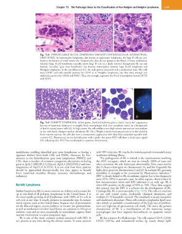
Fig. 73.9 BURKITT LYMPHOMA. At low power, Burkitt lymphoma gives a classic “starry sky” appearance
because of numerous histiocytes or tingible body macrophages with clear cytoplasm (stars), in a background
of darkly stained tumor cells (A). At high power, the cells exhibit a very high mitotic rate and are intermediate
in size with finely clumped nuclear chromatin (B). On a Wright-stained touch preparation or in the blood or
bone marrow aspirate, the cells also have a characteristic appearance with deep blue cytoplasm typically with
vacuoles (C). Fluorescence in situ hybridization with a probe that spans MYC will show a break-apart signal
(D) indicating that MYC has translocated to a partner chromosome.
methylation profiling identified gray zone lymphomas as having a with HIV infection, BL may be the initial acquired immunodeficiency
signature distinct from both CHL and PMBL. However, by fluo- syndrome-defining illness.
rescence in situ hybridization, gray zone lymphomas, PBMCL and The pathogenesis of BL is related to the translocations involving
CHL share a number of common cytogenetic aberrations including the MYC oncogene, which are seen in virtually 100% of cases and
gains at 2p16.1 (REL/BCL11A locus), 9p24.1 (JAK2/PDL2) and rear- often constitute the sole karyotypic abnormality. Most cases involve
rangements of 16p13.13 (CIITA). It is not clear how these patients the IG heavy-chain gene on chromosome 14, and less frequently the
should be approached therapeutically, but they appear to benefit light-chain genes on chromosomes 2 and 22. In endemic BL, genomic
28
from combined modality therapy (systemic chemotherapy and instability is thought to be promoted by Plasmodium infection.
radiation). EBV is closely linked to BL in endemic regions but is less frequently
seen (15%–20%) in sporadic cases. In other regions, characterized by
low socioeconomic status and EBV infection at an early age, BL is
Burkitt Lymphoma often EBV-positive, in the range of 50% to 70%. These data support
the concept that the EBV is a cofactor for the development of BL.
Burkitt lymphoma (BL) is most common in children and accounts for Cytologically, BL is monomorphic (Fig. 73.9). The cells are medium
up to one-third of all pediatric lymphomas in the United States. It is in size with round nuclei, moderately clumped chromatin, and
the most rapidly growing of all lymphomas, with 100% of the cells in multiple (2–5) basophilic nucleoli. The cytoplasm is deeply basophilic
cell cycle at any time. It usually presents in extranodal sites. In nonen- and moderately abundant. These cells contain cytoplasmic lipid vacu-
demic regions, such as the United States, frequent sites of presentation oles, which are probably a manifestation of the high rate of prolifera-
are the ileocecal region, ovaries, kidneys, or breasts. Jaw presentations, tion and high rate of spontaneous cell death. The starry sky pattern
as well as involvement of other facial bones, are common in African or characteristic of BL is a manifestation of the numerous benign
endemic cases and are seen occasionally in nonendemic regions. Bone macrophages that have ingested karyorrhectic or apoptotic tumor
marrow involvement is a poor prognostic sign. cells.
BL is one of the more common tumors associated with HIV. It BL has a mature B cell phenotype. The cells express CD19, CD20,
can present at any time during the clinical course. In some patients CD22, CD79a, and monoclonal surface Ig, nearly always IgM.