
Page 1350 - Hematology_ Basic Principles and Practice ( PDFDrive )
P. 1350
1196 Part VII Hematologic Malignancies
A A
B B C C D D E
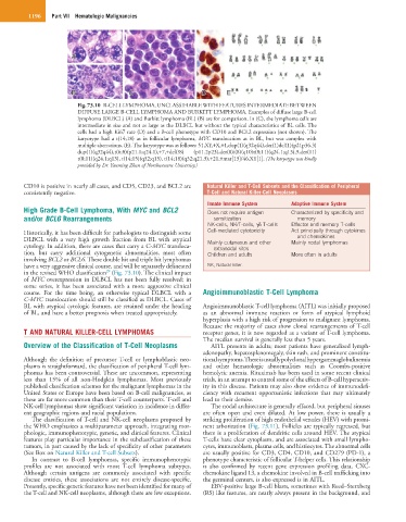
Fig. 73.10 B-CELL LYMPHOMA, UNCLASSIFIABLE WITH FEATURES INTERMEDIATE BETWEEN
DIFFUSE LARGE B-CELL LYMPHOMA AND BURKITT LYMPHOMA. Examples of diffuse large B-cell
lymphoma (DLBCL) (A) and Burkitt lymphoma (BL) (B) are for comparison. In (C), the lymphoma cells are
intermediate in size and not as large as the DLBCL but without the typical characteristics of BL cells. The
cells had a high Ki67 rate (D) and a B-cell phenotype with CD10 and BCL2 expression (not shown). The
karyotype had a t(14;18) as in follicular lymphoma, MYC translocation as in BL, but was complex with
multiple aberrations. (E). The karyotype was as follows: 51,XY,+X,+1,dup(1)(q32q44),der(1)del(1)(p21p36.3)
dup(1)(q32q44),t(6;8)(p21.1;q24.1),+7,+del(?8) (p11.2p23),der(8)i(8)(q10)t(8;11)(q24.1;q13),9,der(11)
t(8;11)(q24.1;q13), t(14;15)(q32;q15), t(14;18)(q32;q21.3),+21,+mar[13]/46,XY[1]. (The karyotype was kindly
provided by Dr. Yanming Zhan of Northwestern University.)
CD10 is positive in nearly all cases, and CD5, CD23, and BCL2 are Natural Killer and T-Cell Subsets and the Classification of Peripheral
consistently negative. T-Cell and Natural Killer-Cell Neoplasms
Innate Immune System Adaptive Immune System
High Grade B-Cell Lymphoma, With MYC and BCL2 Does not require antigen Characterized by specificity and
and/or BCL6 Rearrangements sensitization memory
NK-cells, NK/T-cells, γδ T-cells Effector and memory T-cells
Historically, it has been difficult for pathologists to distinguish some Cell-mediated cytotoxicity Act principally through cytokines
and chemokines
DLBCL with a very high growth fraction from BL with atypical Mainly cutaneous and other Mainly nodal lymphomas
cytology. In addition, there are cases that carry a C-MYC transloca- extranodal sites
tion, but carry additional cytogenetic abnormalities, most often Children and adults More often in adults
involving BCL2 or BCL6. These double hit and triple hit lymphomas
have a very aggressive clinical course, and will be separately delineated NK, Natural killer.
29
in the revised WHO classification (Fig. 73.10). The clinical impact
of MYC overexpression in DLBCL has not been fully resolved; in
some series, it has been associated with a more aggressive clinical
course. For the time being, an otherwise typical DLBCL with a Angioimmunoblastic T-Cell Lymphoma
C-MYC translocation should still be classified as DLBCL. Cases of
BL with atypical cytologic features, are retained under the heading Angioimmunoblastic T-cell lymphoma (AITL) was initially proposed
of BL, and have a better prognosis when treated appropriately. as an abnormal immune reaction or form of atypical lymphoid
hyperplasia with a high risk of progression to malignant lymphoma.
Because the majority of cases show clonal rearrangements of T-cell
T AND NATURAL KILLER-CELL LYMPHOMAS receptor genes, it is now regarded as a variant of T-cell lymphoma.
The median survival is generally less than 5 years.
Overview of the Classification of T-Cell Neoplasms AITL presents in adults; most patients have generalized lymph-
adenopathy, hepatosplenomegaly, skin rash, and prominent constitu-
Although the definition of precursor T-cell or lymphoblastic neo- tional symptoms. There is usually polyclonal hypergammaglobulinemia
plasms is straightforward, the classification of peripheral T-cell lym- and other hematologic abnormalities such as Coombs-positive
phomas has been controversial. These are uncommon, representing hemolytic anemia. Rituximab has been used in some recent clinical
less than 15% of all non-Hodgkin lymphomas. Most previously trials, in an attempt to control some of the effects of B-cell hyperactiv-
published classification schemes for the malignant lymphomas in the ity in this disease. Patients may also show evidence of immunodefi-
United States or Europe have been based on B-cell malignancies, as ciency with recurrent opportunistic infections that may ultimately
these are far more common than their T-cell counterparts. T-cell and lead to their demise.
NK-cell lymphomas show significant variation in incidence in differ- The nodal architecture is generally effaced, but peripheral sinuses
ent geographic regions and racial populations. are often open and even dilated. At low power, there is usually a
The classification of T-cell and NK-cell neoplasms proposed by striking proliferation of high endothelial venules (HEV) with promi-
the WHO emphasizes a multiparameter approach, integrating mor- nent arborization (Fig. 73.11). Follicles are typically regressed, but
phologic, immunophenotypic, genetic, and clinical features. Clinical there is a proliferation of dendritic cells around HEV. The atypical
features play particular importance in the subclassification of these T-cells have clear cytoplasm, and are associated with small lympho-
tumors, in part caused by the lack of specificity of other parameters cytes, immunoblasts, plasma cells, and histiocytes. The abnormal cells
(See Box on Natural Killer and T-cell Subsets). are usually positive for CD3, CD4, CD10, and CD279 (PD-1), a
In contrast to B-cell lymphomas, specific immunophenotypic phenotype characteristic of follicular T-helper cells. This relationship
profiles are not associated with most T-cell lymphoma subtypes. is also confirmed by recent gene expression profiling data. CXC-
Although certain antigens are commonly associated with specific chemokine ligand 13, a chemokine involved in B-cell trafficking into
disease entities, these associations are not entirely disease-specific. the germinal centers, is also expressed is in AITL.
Presently, specific genetic features have not been identified for many of EBV-positive large B-cell blasts, sometimes with Reed–Sternberg
the T-cell and NK-cell neoplasms, although there are few exceptions. (RS) like features, are nearly always present in the background, and