
Page 1352 - Hematology_ Basic Principles and Practice ( PDFDrive )
P. 1352
1198 Part VII Hematologic Malignancies
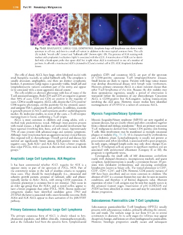
A A B C D
Fig. 73.12 ANAPLASTIC LARGE CELL LYMPHOMA. Anaplastic large cell lymphoma can show a wide
spectrum in cell size, and there is a small cell variant in addition to the more typical common form. The cells
(A) include “wreath cells” (center) and “hallmark cells” (bottom right). (B), The presence of ALK staining with
nuclear and cytoplasmic localization (right) is associated with the t(2;5). A translocation of ALK can be identi-
fied with a break-apart probe that spans ALK but is split when ALK is translocated to one of a number of
partners. A cell with a translocated ALK is pictured in (C) and a normal cell in (D). ALK, Anaplastic lymphoma
kinase.
The cells of classic ALCL have large, often lobulated nuclei with papulosis (LYP) and cutaneous ALCL are part of the spectrum
small basophilic nucleoli, so called hallmark cells. The cytoplasm is of CD30-positive cutaneous T-cell lymphoproliferative diseases.
usually abundant, amphophilic, and there are distinct cytoplasmic Small lesions are likely to regress. Patients with large tumor masses
borders. A prominent Golgi region is generally visible. Small cell and may develop disseminated disease with lymph node involvement.
lymphohistiocytic variants constitute part of the entity, and appear However, primary cutaneous ALCL is a more indolent disease than
to be associated with a more aggressive clinical course. other T-cell lymphomas of the skin. Because the skin nodules may
The cells exhibit an aberrant phenotype with loss of many of the show spontaneous regression, usually a period of observation is
T-cell associated antigens. Both CD3 and CD5 are negative in greater warranted before the institution of any chemotherapy. Cutaneous
than 50% of cases. CD2 and CD4 are positive in the majority of ALCL is CD30-positive but ALK-negative, lacking translocations
cases. CD8 is usually negative. ALCL cells, despite the CD4-positive/ involving the ALK gene. However, recent studies have identified
CD8-negative phenotype, exhibit positivity for the cytotoxic associ- rearrangements of DUSP22 in a subset of cutaneous ALCL.
ated antigens TIA-1, granzyme B, and perforin. In addition, clusterin
is generally present in ALCL and represents another useful diagnostic
marker. By molecular studies, in most of the cases, a T-cell receptor Mycosis Fungoides/Sézary Syndrome
rearrangement is found, confirming a T-cell origin.
ALCL is most common in children and young adults, with a Mycosis fungoides/Sézary syndrome (MF/SS) are now regarded as
marked male predominance noted. Although most patients present separate diseases, but are closely related and often considered together
32
with nodal disease, a high incidence of extranodal involvement has from a clinical and biologic standpoint. Both are primary cutaneous
been reported (involving skin, bone, and soft tissue). Approximately T-cell malignancies derived from mature CD4 positive skin-homing
75% of cases present with advanced-stage and systemic symptoms. T cells. Skin involvement may be manifested as multiple cutaneous
Although these lymphomas have an aggressive natural clinical history, plaques or nodules (Fig. 73.13). SS is characterized by erythroderma
they respond well to chemotherapy. Overall survival and disease-free and a leukemic phase. Lymphadenopathy is usually not present at
survival are significantly better among ALK-positive versus ALK- presentation and, when identified, is associated with a poor prognosis.
negative cases. Both ALK+ and ALK ALCL have a better prognosis In early stages, enlarged lymph nodes may only show changes (Cat-
than other PTCLs, with a plateau in the survival curve seen in both egory I). If malignant cells are present in significant numbers and are
groups. 31 associated with architectural effacement (Category II or III), the
prognosis is significantly worse.
Cytologically, the small cells of MF demonstrate cerebriform
Anaplastic Large Cell Lymphoma, ALK-Negative nuclei with clumped chromatin, inconspicuous nucleoli, and sparse
cytoplasm. Epidermotropism is usually a prominent feature. SS pre-
It has been controversial whether ALCL negative for ALK is a sents with exfoliative erythroderma and circulating cerebriform
+
separate entity or part of the spectrum of PTCL, NOS. Part of lymphocytes known as Sezary cells. The typical phenotype is CD2 ,
+
+
+
the controversy relates to the lack of absolute criteria to recognize CD3 , CD5 , CD4 , and CD8. However, CD8-positive variants of
these cases. They should be morphologically (i.e., sinusoidal and MF have been described, and are more common in children. The
cohesive growth pattern, presence of hallmark cells) and pheno- absence of CD7 is a constant feature but may also be seen in reactive
typically similar to ALK+ ALCL, with strong CD30 expression. A conditions, and therefore is of limited diagnostic value. Aberrant
cytotoxic phenotype is common but is not essential. They occur in expression of other T-cell antigens may be seen but mainly occurs in
an older age group than the ALK+, and as noted earlier, appear to the advanced (tumor) stages. Inactivation of p16 (CDKN2A) and
have a better prognosis than other PTCL, NOS. Recent molecular/ PTEN has been identified in some cases and may be associated with
cytogenetic studies have identified candidate genes TP63 and disease progression.
DUSP22; the latter is associated with a more favorable outcome. Both
ALK+ and ALK ALCL appear to share activation of the JAK/STAT
pathway. Subcutaneous Panniculitis-Like T-Cell Lymphoma
Subcutaneous panniculitis-like T-cell lymphoma (SPTCL) usually
Primary Cutaneous Anaplastic Large Cell Lymphoma presents with subcutaneous nodules, primarily affecting the extremi-
ties and trunk. The nodules range in size from 0.5 cm to several
The primary cutaneous form of ALCL is closely related to lym- centimeters in diameter. In its early stages the infiltrate may appear
phomatoid papulosis, and differs clinically, immunophenotypically, deceptively benign, and lesions are often misdiagnosed as panniculitis.
and at the molecular level from the systemic form. Lymphomatoid However, histologic progression usually occurs and subsequent