
Page 1394 - Hematology_ Basic Principles and Practice ( PDFDrive )
P. 1394
1240 Part VII Hematologic Malignancies
cyclin D1 p53
A B C D E
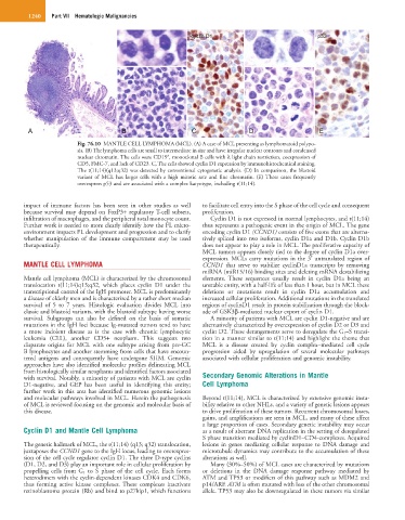
Fig. 76.10 MANTLE CELL LYMPHOMA (MCL). (A) A case of MCL presenting as lymphomatoid polypo-
sis. (B) The lymphoma cells are small to intermediate in size and have irregular nuclear contours and condensed
+
nuclear chromatin. The cells were CD19 , monoclonal B cells with λ light chain restriction, coexpression of
CD5, FMC-7, and lack of CD23. C, The cells showed cyclin D1 expression by immunohistochemical staining.
The t(11;14)(q13;q32) was detected by conventional cytogenetic analysis. (D) In comparison, the blastoid
variant of MCL has larger cells with a high mitotic rate and fine chromatin. (E) These cases frequently
overexpress p53 and are associated with a complex karyotype, including t(11;14).
impact of immune factors has been seen in other studies as well to facilitate cell entry into the S phase of the cell cycle and consequent
because survival may depend on FoxP3+ regulatory T-cell subsets, proliferation.
infiltration of macrophages, and the peripheral total monocyte count. Cyclin D1 is not expressed in normal lymphocytes, and t(11;14)
Further work is needed to more clearly identify how the FL micro- thus represents a pathogenic event in the origin of MCL. The gene
environment impacts FL development and progression and to clarify encoding cyclin D1 (CCND1) consists of five exons that are alterna-
whether manipulation of the immune compartment may be used tively spliced into two isoforms, cyclin D1a and D1b. Cyclin D1b
therapeutically. does not appear to play a role in MCL. The proliferative capacity of
MCL tumors appears closely tied to the degree of cyclin D1a over-
expression. MCLs carry mutations in the 3′ untranslated region of
MANTLE CELL LYMPHOMA CCND1 that serve to stabilize cyclinD1a transcripts by removing
miRNA (miR15/16) binding sites and deleting mRNA destabilizing
Mantle cell lymphoma (MCL) is characterized by the chromosomal elements. These sequences usually result in cyclin D1a being an
translocation t(11;14)q13;q32, which places cyclin D1 under the unstable entity, with a half-life of less than 1 hour, but in MCL these
transcriptional control of the IgH promoter. MCL is predominantly deletions or mutations result in cyclin D1a accumulation and
a disease of elderly men and is characterized by a rather short median increased cellular proliferation. Additional mutations in the translated
survival of 5 to 7 years. Histologic evaluation divides MCL into regions of cyclinD1 result in protein stabilization through the block-
classic and blastoid variants, with the blastoid subtype having worse ade of GSK3β-mediated nuclear export of cyclin D1.
survival. Subgroups can also be defined on the basis of somatic A minority of patients with MCL are cyclin D1-negative and are
mutations in the IgH loci because Ig-mutated tumors tend to have alternatively characterized by overexpression of cyclin D2 or D3 and
a more indolent disease as is the case with chronic lymphocytic cyclin D2. These derangements serve to deregulate the G 1 –S transi-
leukemia (CLL), another CD5+ neoplasm. This suggests two tion in a manner similar to t(11;14) and highlight the theme that
disparate origins for MCL with one subtype arising from pre-GC MCL is a disease created by cyclin complex–mediated cell cycle
B lymphocytes and another stemming from cells that have encoun- progression aided by upregulation of several molecular pathways
tered antigens and consequently have undergone SHM. Genomic associated with cellular proliferation and genomic instability.
approaches have also identified molecular profiles delineating MCL
from histologically similar neoplasms and identified factors associated
with survival. Notably, a minority of patients with MCL are cyclin Secondary Genomic Alterations in Mantle
D1-negative, and GEP has been useful in identifying this entity; Cell Lymphoma
further work in this area has identified numerous genomic lesions
and molecular pathways involved in MCL. Herein the pathogenesis Beyond t(11;14), MCL is characterized by extensive genomic insta-
of MCL is reviewed focusing on the genomic and molecular basis of bility relative to other NHLs, and a variety of genetic lesions appears
this disease. to drive proliferation of these tumors. Recurrent chromosomal losses,
gains, and amplifications are seen in MCL, and many of these affect
a large proportion of cases. Secondary genetic instability may occur
Cyclin D1 and Mantle Cell Lymphoma as a result of aberrant DNA replication in the setting of deregulated
S phase transition mediated by cyclinD1–CD4-complexes. Acquired
The genetic hallmark of MCL, the t(11;14) (q13; q32) translocation, lesions in genes mediating cellular response to DNA damage and
juxtaposes the CCND1 gene to the IgH locus, leading to overexpres- microtubule dynamics may contribute to the accumulation of these
sion of the cell cycle regulator cyclin D1. The three D-type cyclins alterations as well.
(D1, D2, and D3) play an important role in cellular proliferation by Many (30%–50%) of MCL cases are characterized by mutations
propelling cells from G 1 to S phase of the cell cycle. Each forms or deletions in the DNA damage response pathway mediated by
heterodimers with the cyclin-dependent kinases CDK4 and CDK6, ATM and TP53 or modifiers of this pathway such as MDM2 and
thus forming active kinase complexes. These complexes inactivate p14/ARF. ATM is often mutated with loss of the other chromosomal
retinoblastoma protein (Rb) and bind to p27kip1, which functions allele. TP53 may also be downregulated in these tumors via similar