
Page 1395 - Hematology_ Basic Principles and Practice ( PDFDrive )
P. 1395
Chapter 76 Origin of Non-Hodgkin Lymphoma 1241
mechanisms, and overexpression of TP53-negative regulators MDM2 metabolism and are tightly intertwined with abnormalities in cell
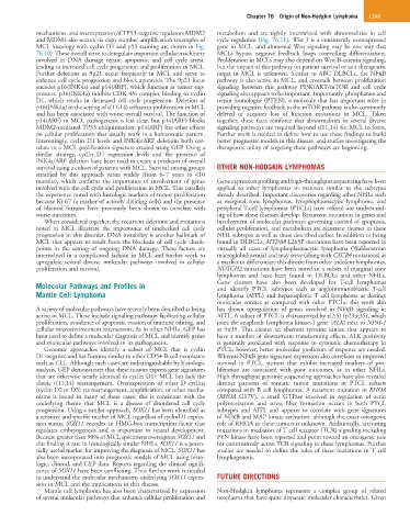
and MDM4 also occurs via copy number amplification (examples of cycle regulation (Fig. 76.11). Wnt 3 is a consistently overexpressed
MCL histology with cyclin D1 and p53 staining are shown in Fig. gene in MCL, and abnormal Wnt signaling may be one way that
76.10). These overall serve to deregulate important cellular machinery MCLs bypass negative feedback loops controlling differentiation.
involved in DNA damage repair, apoptosis, and cell cycle arrest, Proliferation in MCLs may also depend on Wnt-B-catenin signaling,
leading to increased cell cycle progression and proliferation in MCL. but the impact of this pathway on patient survival or as a therapeutic
Further deletions at 9p21 occur frequently in MCL and serve to target in MCL is unknown. Similar to ABC DLBCLs, the NFκB
enhance cell cycle progression and block apoptosis. The 9p21 locus pathway is also active in MCL, and crosstalk between proliferation
encodes p16(INK4a) and p14(ARF), which function as tumor sup- signaling between this pathway PI3K/AKT/mTOR and cell cycle
pressors. p16(INK4a) inhibits CDK 4/6 complex binding to cyclin signaling also appears to be important. Importantly, phosphatase and
D1, which results in decreased cell cycle progression. Deletion of tensin homologue (PTEN), a molecule that has important roles in
p16(INK4a) in the setting of t(11;14) enhances proliferation in MCL providing negative feedback to the mTOR pathway is also commonly
and has been associated with worse overall survival. The function of deleted or acquires loss of function mutations in MCL. Taken
p14(ARF) in MCL pathogenesis is less clear, but p14(ARF) blocks together, these facts reinforce that abnormalities in several diverse
MDM2-mediated TP53 ubiquitination; p14(ARF) has other effects signaling pathways are required beyond t(11;14) for MCL to form.
on cellular proliferation that usually work in a homeostatic pattern. Further work is needed to define how to use these findings to build
Interestingly, cyclin D1 levels and INK4a/ARF deletions both cor- better prognostic models in this disease, and studies investigating the
relate to a MCL proliferation signature created using GEP. Using a therapeutic utility of targeting these pathways are beginning.
similar strategy, cyclin D1 expression levels and the presence of
INK4a/ARF deletion have been used to create a predictor of overall
survival using a cohort of patients with MCL. Survival among groups OTHER NON-HODGKIN LYMPHOMAS
stratified by this approach varies widely (from 6–7 years to <10
months), which confirms the importance of involvement of genes Gene expression profiling and high-throughput sequencing have been
involved with the cell cycle and proliferation in MCL. This parallels applied to other lymphomas in manners similar to the subtypes
the experience noted with histologic markers of tumor proliferation already described. Important discoveries regarding other NHLs such
because Ki-67 (a marker of actively dividing cells) and the presence as marginal zone lymphomas, lymphoplasmacytic lymphoma, and
of blastoid features have previously been shown to correlate with peripheral T-cell lymphomas (PTCLs) have refined our understand-
worse outcomes. ing of how these diseases develop. Recurrent mutations in genes and
When considered together, the recurrent deletions and mutations involvement of molecular pathways governing control of apoptosis,
noted in MCL illustrate the importance of unchecked cell cycle cellular proliferation, and metabolism are recurrent themes in these
progression in this disorder. DNA instability is another hallmark of NHL subtypes as well as those described earlier. In addition to being
MCL that appears to result from the blockade of cell cycle check- found in DLBCLs, MYD88 L265P mutations have been reported in
points in the setting of ongoing DNA damage. These factors are virtually all cases of lymphoplasmacytic lymphoma (Waldenström
intertwined in a complicated fashion in MCL and further work to macroglobulinemia) and may serve (along with CXCR4 mutations) as
upregulate several diverse molecular pathways involved in cellular a marker to differentiate this disease from other indolent lymphomas.
proliferation and survival. NOTCH2 mutations have been noted in a subset of marginal zone
lymphomas and have been found in DLBCLs and other NHLs.
Molecular Pathways and Profiles in Gene clusters have also been developed for T-cell lymphomas
and identify PTCL subtypes such as angioimmunoblastic T-cell
Mantle Cell Lymphoma lymphoma (AITL) and hepatosplenic T cell lymphoma as distinct
molecular entities as compared with other PTCLs; this work also
A variety of molecular pathways have recently been described as being has shown upregulation of genes involved in NFκB signaling in
active in MCL. These include signaling pathways facilitating cellular AITL. A subset of PTCL is characterized by t(2;5) (p23;q35), which
proliferation, avoidance of apoptosis, evasion of immune editing, and joins the anaplastic lymphoma kinase-1 gene (ALK) next to NPM-1
cellular microenvironment interactions. As in other NHLs, GEP has at 5q35. This creates an aberrant tyrosine kinase that appears to
been used to define a molecular diagnosis of MCL and identify genes have a number of downstream transforming effects. ALK positivity
and molecular pathways involved in its pathogenesis. is generally associated with response to cytotoxic chemotherapy in
Genomic approaches identify a subset of MCL that is cyclin PTCL; however, better molecular predictors of response are needed.
D1-negative and has features similar to other CD5+ B-cell neoplasms Whereas NFκB gene signature expression also correlates to improved
such as CLL. Although such cases are indistinguishable by histologic survival in PTCL, tumors that exhibit increased markers of pro-
analysis, GEP demonstrates that these tumors express gene signatures liferation are associated with poor outcomes, as in other NHLs.
that are otherwise nearly identical to cyclin D1+ MCL but lack the High throughput genomic sequencing approaches have also revealed
classic t(11;14) rearrangement. Overexpression of other D cyclins distinct patterns of somatic tumor mutations in PTCL subsets
(cyclin D2 or D3) via rearrangement, amplification, or other mecha- compared with B cell lymphomas. A recurrent mutation in RHOA
nisms is found in many of these cases; this is consistent with the (RHOA G17V), a small GTPase involved in regulation of actin
underlying theme that MCL is a disease of disordered cell cycle polymerization and stress fiber formation occurs in both PTCL
progression. Using a similar approach, SOX11 has been identified as subtypes and AITL and appears to correlate with gene signatures
a sensitive and specific marker of MCL regardless of cyclinD1 expres- of NFκB and MAP kinase activation, although the exact oncogenic
sion status. SOX11 encodes an HMG-box transcription factor that role of RHOA in these tumors is unknown. Additionally, activating
regulates embryogenesis and is important in neural development. mutations in mediators of T cell receptor (TCR) signaling including
Because greater than 90% of MCL specimens overexpress SOX11 and FYN kinase have been reported and point toward an oncogenic role
this finding is rare in histologically similar NHLs, SOX11 is a poten- for constitutively active TCR signaling in these lymphomas. Further
tially useful marker for improving the diagnosis of MCL. SOX11 has studies are needed to define the roles of these mutations in T cell
also been incorporated into prognostic models of MCL using histo- lymphogenesis.
logic, clinical, and GEP data. Reports regarding the clinical signifi-
cance of SOX11 have been conflicting. Thus further work is needed
to understand the molecular mechanisms underlying SOX11 expres- FUTURE DIRECTIONS
sion in MCL and the implications in this disease.
Mantle cell lymphoma has also been characterized by expression Non-Hodgkin lymphoma represents a complex group of related
of several molecular pathways that enhance cellular proliferation and neoplasms that have quite disparate molecular characteristics. Given