
Page 2136 - Hematology_ Basic Principles and Practice ( PDFDrive )
P. 2136
Chapter 126 Molecular Basis of Blood Coagulation 1897
Thrombin
Fibrinogen vWF
GPIIb/IIIa
PARS(1,4)
GPIb-IX-V
ADP Collagen
P2Y 12
GPIa/IIa
Platelet
Epinephrine activation TXA 2 R
α 2 A-AR
TX synthase
PGG 2 TXA 2
Aspirin
COX-1
Arachidonic acid
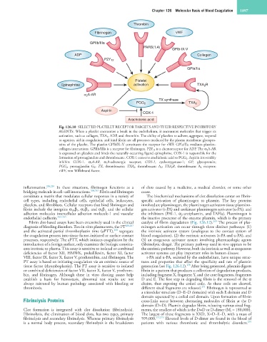
Fig. 126.10 SELECTED PLATELET RECEPTOR TARGETS AND THEIR RESPECTIVE INHIBITORY
AGENTS. When a platelet encounters a break in the endothelium, it encounters molecules that trigger its
activation, such as collagen, TXA 2, ADP, and thrombin. The ability of platelets to adhere, aggregate, respond
to agonists, aid in coagulation, and bind fibrin are all processes mediated by the plasma membrane glycopro-
teins of the platelet. The platelet GPIbIX–V constitutes the receptor for vWF. GPIa/IIa mediates platelet–
collagen interactions. GPIIb/IIIa is a receptor for fibrinogen. P2Y 12 is a chemoreceptor for ADP. The α 2A-AR
is expressed on platelets and binds the naturally occurring ligand epinephrine. COX-1 is responsible for the
formation of prostaglandins and thromboxane. COX-1 converts arachidonic acid to PGG 2. Aspirin irreversibly
inhibits COX-1. α 2A-AR, α 2A-adrenergic receptor; COX-1, cyclooxygenase-1; GP, glycoprotein;
PGG 2, prostaglandin G 2 ; TX, thromboxane; TXA 2, thromboxane A 2; TXA 2R, thromboxane A 2 receptor;
vWF, von Willebrand factor.
inflammation. 258,259 In these situations, fibrinogen functions as a of clots caused by a medicine, a medical disorder, or some other
bridging molecule in cell–cell interactions. 260,261 Fibrin and fibrinogen cause.
constitute a matrix that modulates cellular responses in a variety of The biochemical mechanisms of clot dissolution center on fibrin-
cell types, including endothelial cells, epithelial cells, leukocytes, specific activation of plasminogen to plasmin. The key proteins
platelets, and fibroblasts. Cellular receptors that bind fibrinogen and involved are plasminogen, the plasminogen activators tissue plasmino-
fibrin include the integrins α IIb β 3 , α V β 3 , and α 5 β 1, and the cellular gen activator (t-PA) and urokinase plasminogen activator (u-PA), and
adhesion molecules intercellular adhesion molecule-1 and vascular the inhibitors (PAI-1, α 2 -antiplasmin, and TAFIa). Plasminogen is
endothelial cadherin. 262–265 the inactive precursor of the enzyme plasmin, which is the primary
269
Fibrin clot-based assays have been extensively used in the clinical catalyst of fibrin degradation (Fig. 126.12). The process of plas-
diagnosis of bleeding disorders. Two in vitro plasma tests, the PT 266,267 minogen activation can occur through three distinct pathways: (1)
268
and the activated partial thromboplastin time (aPTT), segregate the intrinsic activator system (analogous to the contact system of
the coagulation process into tissue factor–initiated or surface contact blood coagulation), (2) the extrinsic activators (t-PA and u-PA), and
processes, respectively. The aPTT, which initiates coagulation by the (3) an exogenous activator system involving pharmacologic agents
introduction of a foreign surface, only examines the biologic constitu- (fibrinolytic drugs). The primary pathway used in vivo appears to be
ents intrinsic to plasma. This assay is sensitive to isolated or combined the extrinsic pathway. However, both the intrinsic as well as exogenous
deficiencies of factor XII, HMWK, prekallikrein, factor XI, factor activator systems can play important roles in human disease.
VIII, factor IX, factor X, factor V, prothrombin, and fibrinogen. The t-PA and u-PA, secreted by the endothelium, have unique struc-
PT assay is based on initiating coagulation via an extrinsic source of tures and properties that affect the specificity and rate of plasmin
270
tissue factor (thromboplastin). The PT assay is sensitive to isolated generation (see Fig. 126.12). After being generated, plasmin digests
or combined deficiencies of factor VII, factor X, factor V, prothrom- fibrin in a pattern that produces a collection of degradation products,
bin, and fibrinogen. Although these in vitro clotting assays help including fragment X, fragment Y, and the core fragments, fragments
establish a basis for hemostasis, abnormal test results are not D and E. The first step in degrading fibrin is the removal of the α
always mirrored by human pathology associated with bleeding or chains, thus exposing the coiled coils. As these coils are cleaved,
271
thrombosis. different sized fragments are released. Fibrinogen is represented as
a trinodular structure (D–E–D domains) with each E domain and D
domain separated by a coiled coil domain. Upon formation of fibrin
Fibrinolysis Proteins cross-links occur between alternating molecules of fibrin at the D
domain (D=D). Plasmin degrades fibrin, releasing various sized frag-
Clot formation is integrated with clot dissolution (fibrinolysis). ments, the smallest of which is the D=D or D-dimer (M r = 180,000).
Fibrinolysis, the elimination of blood clots, has two types, primary The largest of these fragments is XXD, X=D–E–D, with a mass of
fibrinolysis and secondary fibrinolysis. Whereas primary fibrinolysis 595,000. 272,273 Elevated levels of D-dimer are found in the blood of
274
is a normal body process, secondary fibrinolysis is the breakdown patients with various thrombotic and thrombolytic disorders.