
Page 2317 - Hematology_ Basic Principles and Practice ( PDFDrive )
P. 2317
Chapter 138 Structure, Biology, and Genetics of von Willebrand Factor 2059
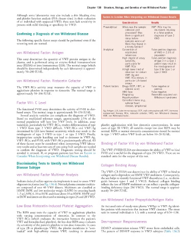
Although some laboratories may also include a skin bleeding time Factors to Consider When Interpreting von Willebrand Disease Results
and platelet function analysis (PFA closure time) in their evaluation
of an individual with suspected VWD, these tests lack sensitivity in Considerations Results
persons with mild bleeding or specificity for VWD.
Preanalytical When was the sample VWF : RCo may be
collected and decreased resulting
Confirming a Diagnosis of von Willebrand Disease processed? Was in a false-positive
there a significant diagnosis of type 2
delay before VWD
The following specific factor assays should be performed even if the samples were run?
screening tests are normal. Were they frozen in
a timely fashion?
Analytical Convention of False-positive diagnosis
von Willebrand Factor: Antigen established of VWD in 2.5% of
references population
This assay determines the quantity of VWF protein antigen in the High degree of assay False-positive diagnosis
plasma, and is performed using an enzyme-linked immunosorbent variability, of type 2 in a type 1
particularly for
patient may result in
assay (ELISA) or latex immunoassay (LIA). The normal range (which VWF : RCo the misdiagnosis of
should be determined independently by each laboratory) is approxi- A high lower limit of type 3 for type 1 or
mately 50–200 IU/dL. detection for certain type 2 VWD
VWF : Ag and
VWF : RCo assays, in
von Willebrand Factor: Ristocetin Cofactor particular LIA-based
assays
The VWF : RCo activity assay measures the capacity of VWF to Patient factors Drugs (OCP, HRT, or False negative or
positive
valproic acid)
agglutinate platelets in response to ristocetin. The normal range is ABO type False negative
approximately 50–200 IU/dL. Pregnancy Reversible acquired von
Hypothyroidism Willebrand syndrome
Comorbid illness (e.g.,
Factor VIII: C Level valvular heart
disease, lymphoma)
The functional FVIII assay determines the activity of FVIII in clot- Ag, Antigen; LIA, latex immunoassay; OCP, oral contraceptive pill; HRT, hormone
based assays. The normal range is approximately 50–150 IU/dL. replacement therapy; RCo, ristocetin cofactor; VWD, von Willebrand disease;
Several analytic variables can complicate the diagnosis of VWD. VWF, von Willebrand factor.
Based on established reference ranges, approximately 2.5% of the
normal population will have low VWF levels. In addition, assay
variability, particularly for VWF : RCo, renders differentiation of type platelet agglutination with low ristocetin concentrations. In some
1 VWD from type 2 VWD difficult. VWF : RCo and VWF : Ag cases of type 2B VWD, all variables except low dose RIPA may be
determined by LIA have limited sensitivity, which may result in the normal. RIPA at normal ristocetin concentrations should be normal
misdiagnosis of type 3 VWD as type 1 or type 2 VWD. Finally, in type 1 VWD unless VWF levels are below 10–20 IU/dL.
inappropriate sample handling can lead to decreases in VWF : Ag,
VWF : RCo, and FVIII, with VWF : RCo predominantly affected. All
of these factors must be considered when interpreting VWF labora- Binding of Factor VIII by von Willebrand Factor
tory results and at least two sets of tests using fresh samples are needed
to confirm the diagnosis of VWD. Diagnostic testing should be The VWF : FVIIIB ELISA test determines the ability of VWF to bind
avoided in stressed, ill, or pregnant patients (see box on Factors to FVIII and is useful for the diagnosis of type 2N VWD. There are no
Consider When Interpreting von Willebrand Disease Results). standard units for the output of this test.
Discriminating Tests to Identify von Willebrand Collagen Binding Assay
Disease Subtype
The VWF : CB ELISA test determines the ability of VWF to bind to
von Willebrand Factor Multimer Analysis collagen and is dependent on HMW VWF multimers. Consequently,
the test helps to identify functional VWF discordance (i.e., to distin-
Sodium dodecyl sulfate-agarose electrophoresis is used to assess VWF guish between types 1 and 2 VWD). Reduced collagen binding
oligomers in plasma (see Fig. 138.4). Normal plasma contains multim- reflects the loss of HMW multimers or can reflect a specific collagen-
ers composed of over 40 VWF dimers. Multimers are classified as binding deficiency (type 2M VWD). The normal range is approxi-
HMW, IMW, and low molecular weight (LMW) by counting bands mately 50–200 IU/dL.
1–5 as LMW, 6–10 as IMW, and those above 10 as HMW. HMW and/
or IMW multimers are decreased or missing in types 2A and 2B VWD.
von Willebrand Factor Propeptide/Antigen Ratio
Low Dose Ristocetin-Induced Platelet Aggregation An increased ratio of steady-state plasma VWFpp to VWF : Ag identi-
fies patients with mutations that increase VWF clearance. The mean
The RIPA assay tests the capacity of VWF to agglutinate platelets ratio in normal individuals is 1.3, with a normal range of 0.54–1.98.
with varying concentrations of ristocetin. In contrast to the
VWF : RCo (which evaluates the interaction between the patient’s
VWF and formalin-fixed platelets), the low dose RIPA assay evaluates Desmopressin Responsiveness
the sensitivity of the patient’s platelets to low-dose ristocetin. In cases
of type 2B or platelet-type VWD, the platelet membrane is “over- DDAVP administration releases VWF stores from endothelial cells.
loaded” with high-affinity mutant VWF, resulting in abnormal The pattern of DDAVP response in VWD subtypes (Table 138.3)