
Page 2314 - Hematology_ Basic Principles and Practice ( PDFDrive )
P. 2314
2056 Part XII Hemostasis and Thrombosis
drugs targeting VWF attenuate arterial thrombosis in animal models. secretion or clearance, which also lead to a VWD phenotype. A third
Their utility in humans is unknown. level of classification denoted by roman numerals (e.g., VWD type
2A IIA) indicates specific phenotypes and is a remnant of an older
classification system that is mainly used in the research setting.
VON WILLEBRAND DISEASE
VWD is caused by deficient or defective plasma VWF and represents von Willebrand Disease Type 1
the most common inherited bleeding disorder. Bleeding symptoms
reflect the defect in primary hemostasis: mucocutaneous bleeding, Type 1 VWD, a quantitative deficiency of VWF, represents approxi-
especially epistaxis and menorrhagia. When FVIII levels are suffi- mately 70% of VWD cases. The VWF is functionally normal without
ciently low, the bleeding phenotype overlaps with that of mild to a specific abnormality in ligand binding sites or a significant decrease
moderate hemophilia; patients may experience joint or muscle bleeds. in HMW multimers. Functional assays of VWF, such as VWF : RCo,
The current VWD classification recognizes three subtypes. Type 1 are decreased in proportion to the decrease in VWF : Ag concentra-
VWD is characterized by quantitative deficiency of VWF, type 2 tion, and the ratio of functional activity as compared with VWF : Ag
VWD is characterized by qualitative defects, and type 3 VWD is is normal (i.e., VWF : RCo/VWF : Ag ratio is >0.6).
characterized by an almost complete absence of VWF. Point mutations, most frequently missense mutations, have been
identified in approximately 65% of individuals with type 1 VWD
and occur throughout the VWF gene. Fully penetrant, dominantly
Epidemiology inherited missense mutations are more often identified when
VWF : Ag and VWF : RCo levels are less than 25 IU/dL. In contrast,
VWD is the most common inherited bleeding disorder. However, incompletely penetrant, dominantly inherited missense mutations,
because VWF levels are variable in the population and symptom such as p.Tyr1584Cys and p.Arg924Gln, are identified in approxi-
severity ranges from infrequent, mild bleeding to frequent or severe mately 50% of individuals whose VWF : Ag and VWF : RCo levels
bleeds, the reported prevalence depends on the diagnostic criteria and are above 25 IU/dL. The extent to which incompletely penetrant
the study population. In two large epidemiologic studies, the preva- VWF mutations contribute to the bleeding phenotype in individuals
lence of VWD was approximately 1% in healthy school-age children with VWF levels of approximately 50 IU/dL is not clear, and genetic
based on low VWF activity, and a personal and family history of analyses in such cases are difficult to interpret.
bleeding symptoms. The prevalence of VWD in individuals who Missense mutations may affect VWF levels by reducing secretion
present to a primary care physician with bleeding symptoms is and/or increasing clearance. The most frequently reported genetic
approximately 0.1%, whereas in patients whose bleeding symptoms mutation is a missense mutation that results in the substitution of
are sufficiently severe to warrant referral to specialized centers, the tyrosine with cysteine at codon 1584 (Y1584C), which is found in
prevalence is 20–113 per million. 10% to 20% of type 1 VWD patients. Intracellular retention is a
common mechanism for type 1 VWD pathogenicity and can result
from missense mutations in various VWF domains. Haploinsuffi-
Classification and Pathophysiology ciency from a heterozygous null allele results in reduced VWF
expression in a small proportion of cases. A common heterozygous
The 2006 ISTH VWD classification relies on the VWF protein in-frame large deletion of exons 4–5 was reported in a cohort of type
phenotype, which in turn often reflects the underlying pathophysiol- 1 VWD patients in the United Kingdom, and this and similar partial
ogy and has implications for treatment. Type 1 VWD is a partial gene deletions may contribute to the spectrum of mutations in a
quantitative deficiency; type 2 (with four subtypes: 2A, 2B, 2M, and minority of cases. A well-described pathophysiologic mechanism for
2N) is a qualitative defect; and type 3 is a virtual deficiency of VWF VWD type 1 is increased VWF clearance, referred to as type 1C (C
(Table 138.1). The diagnosis and categorization of VWD into a type for increased clearance), although this designation is not included in
can be achieved with widely available laboratory testing. However, the ISTH classification. Patients typically have very low VWF levels,
the differentiation among type 2 subtypes may require referral to a an increased vWFpp/VWF : Ag ratio, and a marked but short-lived
specialized laboratory. The current classification does not incorporate response to DDAVP. Of note, the half-life of VWF/FVIII concen-
genotypic data: the diagnosis of VWD is not limited to individuals trates is normal in these individuals. Missense mutations mainly
with mutations within the VWF gene. VWF mutations may not be occur in the D3 domain and reduce the half-life of VWF up to
identified in VWD patients because of the complexity of the VWF 15-fold. R1205H, which is known as the “Vicenza” variant, is the
gene or because of mutations in other genes, such as those affecting most common, most severe, and best characterized of these muta-
tions. Because of the transient response to DDAVP, the utility of this
medication for treatment of major bleeds is questioned in this sub-
group of patients.
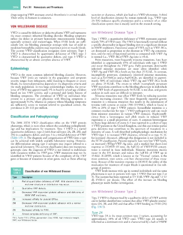
TABLE Classification of von Willebrand Disease VWF levels increase with age in normal individuals and the same
138.1 phenomena is seen in patients with type 1 VWD (but not type 2 or
Type Description 3). The increase has been reported as 3.5 U/dL VWF : Ag and 7.1 U/
dL FVIII : C per decade. The effect of this increase on bleeding
1 Partial quantitative deficiency of VWF. Mild abnormalities in phenotype needs further investigation.
multimer structure or distribution may occur.
2 Qualitative VWF defects.
2A Decreased VWF-dependent platelet adhesion and deficiency of von Willebrand Disease Type 2
HMW VWF multimers.
Type 2 VWD is characterized by a qualitative defect of VWF activity
2B Increased affinity for platelet GPIbα.
and is further classified into variants that affect VWF-platelet interac-
2M Decreased VWF-dependent platelet adhesion with a normal tions (2A, 2B, and 2M) and that affect VWF binding to FVIII (2N)
multimer distribution. (Fig. 138.5).
2N Decreased affinity for FVIII.
3 Almost complete deficiency of VWF. Type 2A
VWD type 2A is the most common type 2 variant, accounting for
FVIII, Factor FVIII; GPIbα, glycoprotein 1bα; HMW, high molecular weight; approximately 10% of all VWD cases. VWD type 2A usually is
VWF, von Willebrand factor.
inherited as a dominant trait and is characterized by a lack of HMW