
Page 2315 - Hematology_ Basic Principles and Practice ( PDFDrive )
P. 2315
Chapter 138 Structure, Biology, and Genetics of von Willebrand Factor 2057
Gp1b
FVIII Collagen Collagen Dimerization
Multimerization
5′ UT D1 D2 D’ D3 A1 A2 A3 D4 B C1 C2
Exon No. 12 3–10 11–17 18–20 20–28 28 28 28–32 35–39 40–4244–48 49–52
Type 2N Types Type 2A Type 2A
2B and 2M
Propeptide Mature VWF monomer
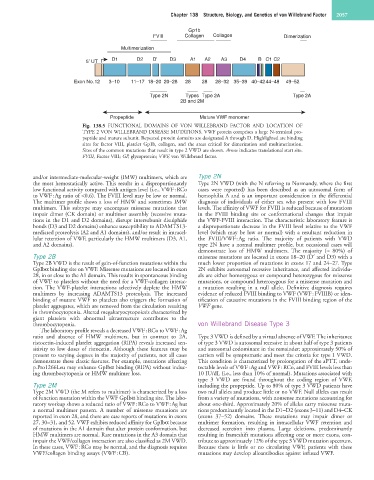
Fig. 138.5 FUNCTIONAL DOMAINS OF VON WILLEBRAND FACTOR AND LOCATION OF
TYPE 2 VON WILLEBRAND DISEASE MUTATIONS. VWF protein comprises a large N-terminal pro-
peptide and mature subunit. Repeated protein domains are designated A through D. Highlighted are binding
sites for factor VIII, platelet Gp1b, collagen, and the areas critical for dimerization and multimerization.
Sites of the common mutations that result in type 2 VWD are shown. Arrow indicates translational start site.
FVIII, Factor VIII; GP, glycoprotein; VWF, von Willebrand factor.
and/or intermediate-molecular-weight (IMW) multimers, which are Type 2N
the most hemostatically active. This results in a disproportionately Type 2N VWD (with the N referring to Normandy, where the first
low functional activity compared with antigen level (i.e., VWF : RCo cases were reported) has been described as an autosomal form of
to VWF : Ag ratio of <0.6). The FVIII level may be low or normal. hemophilia A and is an important consideration in the differential
The multimer profile shows a loss of HMW and sometimes IMW diagnosis of individuals of either sex who present with low FVIII
multimers. This subtype may encompass missense mutations that levels. The affinity of VWF for FVIII is reduced because of mutations
impair dimer (CK domain) or multimer assembly (recessive muta- in the FVIII binding site or conformational changes that impair
tions in the D1 and D2 domains), disrupt intersubunit disulphide the VWF-FVIII interaction. The characteristic laboratory feature is
bonds (D3 and D2 domains) enhance susceptibility to ADAMTS13- a disproportionate decrease in the FVIII level relative to the VWF
mediated proteolysis (A2 and A1 domains), and/or result in intracel- level (which may be low or normal) with a resultant reduction in
lular retention of VWF, particularly the HMW multimers (D3, A1, the FVIII/VWF : Ag ratio. The majority of patients with VWD
and A2 domains). type 2N have a normal multimer profile, but occasional cases will
demonstrate loss of HMW multimers. The majority (≈ 80%) of
Type 2B missense mutations are located in exons 18–20 (D′ and D3) with a
Type 2B VWD is the result of gain-of-function mutations within the much lower proportion of mutations in exons 17 and 24–27. Type
GpIbα binding site on VWF. Missense mutations are located in exon 2N exhibits autosomal recessive inheritance, and affected individu-
28, in or close to the A1 domain. This results in spontaneous binding als are either homozygous or compound heterozygous for missense
of VWF to platelets without the need for a VWF-collagen interac- mutations, or compound heterozygous for a missense mutation and
tion. The VWF-platelet interactions selectively deplete the HMW a mutation resulting in a null allele. Definitive diagnosis requires
multimers by increasing ADAMTS13 proteolysis. The increased evidence of reduced FVIII binding to VWF (VWF : FVIIIB) or iden-
binding of mutant VWF to platelets also triggers the formation of tification of causative mutations in the FVIII binding region of the
platelet aggregates, which are removed from the circulation resulting VWF gene.
in thrombocytopenia. Altered megakaryocytopoiesis characterized by
giant platelets with abnormal ultrastructure contributes to the
thrombocytopenia. von Willebrand Disease Type 3
The laboratory profile reveals a decreased VWF : RCo to VWF : Ag
ratio and absence of HMW multimers, but in contrast to 2A, Type 3 VWD is defined by a virtual absence of VWF. The inheritance
ristocetin-induced platelet aggregation (RIPA) reveals increased sen- of type 3 VWD is autosomal recessive in about half of type 3 patients
sitivity to low doses of ristocetin. Although these features may be and autosomal codominant in the remainder: approximately 50% of
present to varying degrees in the majority of patients, not all cases carriers will be symptomatic and meet the criteria for type 1 VWD.
demonstrate these classic features. For example, mutations affecting This condition is characterized by prolongation of the aPTT, unde-
p.Pro1266Leu may enhance GpIbα binding (RIPA) without induc- tectable levels of VWF : Ag and VWF : RCo, and FVIII levels less than
ing thrombocytopenia or HMW multimer loss. 10 IU/dL (i.e., less than 10% of normal). Mutations associated with
type 3 VWD are found throughout the coding region of VWF,
Type 2M including the propeptide. Up to 80% of type 3 VWD patients have
Type 2M VWD (the M refers to multimer) is characterized by a loss two null alleles and produce little or no VWF. Null alleles can result
of function mutation within the VWF GpIbα binding site. The labo- from a variety of mutations, with nonsense mutations accounting for
ratory workup shows a reduced ratio of VWF : RCo to VWF : Ag but about one-third. Approximately 20% of alleles carry missense muta-
a normal multimer pattern. A number of missense mutations are tions predominantly located in the D1–D2 (exons 3–11) and D4–CK
reported in exon 28, and there are case reports of mutations in exons (exons 37–52) domains. These mutations may impair dimer or
27, 30–31, and 52. VWF exhibits reduced affinity for GpIbα because multimer formation, resulting in intracellular VWF retention and
of mutations in the A1 domain that alter protein conformation, but decreased secretion into plasma. Large deletions, predominantly
HMW multimers are normal. Rare mutations in the A3 domain that resulting in frameshift mutations affecting one or more exons, con-
impair the VWF/collagen interaction are also classified as 2M VWD. tribute to approximately 12% of the type 3 VWD mutation spectrum.
In these cases, VWF : RCo may be normal, and the diagnosis requires Because there is little or no circulating VWF, patients with these
VWF/collagen binding assays (VWF : CB). mutations may develop alloantibodies against infused VWF.