
Page 2316 - Hematology_ Basic Principles and Practice ( PDFDrive )
P. 2316
2058 Part XII Hemostasis and Thrombosis
Clinical Manifestations Type 1 VWD accounts for up to 70% of VWD. It typically
manifests as mild mucocutaneous bleeding; however, symptoms may
The bleeding history depends on disease severity; type 3 VWD is be more severe if VWF levels are below 15 IU/dL. Epistaxis and
often diagnosed early in life, whereas mild type 1 VWD may not be bruising are common symptoms in children. Menorrhagia is the most
diagnosed until adulthood. Individuals with VWD primarily com- common finding in women of reproductive age.
plain of excessive mucocutaneous bleeding, such as spontaneous, Type 2 VWD accounts for about 25% of all VWD. The relative
recurrent epistaxis, and prolonged bleeding after dental cleaning or frequency of the subtypes is 2A > 2N > 2M > 2B in European popula-
extraction. In addition, prolonged or excessive bleeding after surgery tions. Individuals with type 2A, 2B, and 2M VWD usually present
or trauma is often reported. Affected females frequently experience with mild to moderate mucocutaneous bleeding, but bleeding epi-
menorrhagia from the time of menarche, and can have prolonged or sodes can be severe, particularly when VWF : RCo is very low or
excessive bleeding after childbirth. Musculoskeletal bleeding is absent. In many patients with type 2B VWD, thrombocytopenia can
unusual, except in type 2N or type 3 VWD when the FVIII : C level develop or worsen with infection, surgery, pregnancy, or treatment
may be below 10 IU/dL. Bleeding assessment tools (BATs) help to with DDAVP. The symptoms of type 2N VWD are similar to those
standardize and quantify the bleeding history. BATs are helpful for seen in mild hemophilia A (see Chapter 135) because both disorders
making a VWD diagnosis. In addition, high scores are predictive of are associated with reduced levels of FVIII : C.
the risk of future bleeding. (see Chapter 128) Type 3 VWD is the rarest subtype and accounts for less than 5%
of VWD. Prevalence estimates range from 0.55 to 6 per million, with
higher rates seen with consanguineous marriage. Type 3 VWD mani-
Example of the Presentation and Impact of Type 1 von fests with severe bleeding, including excessive mucocutaneous bleed-
Willebrand Disease ing and musculoskeletal bleeding.
J.C., a 26-year-old female, presents with delayed postpartum hemor-
rhage (PPH) after the delivery of her first child. The pregnancy and Penetrance
delivery were unremarkable. One week postpartum, the patient expe-
rienced heavy bleeding, and required a dilation and curettage. At the In autosomal dominant type 1 VWD, mutations resulting in plasma
same time, she was started on tranexamic acid, which was continued VWF level less than 25 IU/dL are often fully penetrant, whereas
for 2 weeks. By approximately 3 weeks postpartum, there was com- those resulting in higher VWF levels are often incompletely penetrant.
plete cessation of bleeding. A bleeding history was significant for the
following: excessive postsurgical bleeding following tonsillectomy and Mutations responsible for autosomal dominant types of VWD (2A,
adenoidectomy, which required red blood cell transfusion; excessive 2B, and 2M) are often fully penetrant. Thus in contrast to the vari-
bleeding postdental extractions, requiring consultation with the dentist ably positive family histories in patients with type 1 VWD, those
and repacking; and menorrhagia from the time of menarche, which with type 2 VWD usually have a positive family history.
had been well controlled with the combined oral contraceptive pill.
There was no family history of VWD. Investigations revealed VWF : Ag
0.40 IU/dL, VWF : RCo 0 : 39 IU/dL, and FVIII : C 0.60 IU/dL. Multimer Laboratory Investigations
gel was unremarkable. A DDAVP challenge demonstrated that the
patient was a responder. She became pregnant again 4 years later. At The laboratory evaluation for VWD involves a battery of qualitative
the time of delivery she was treated with tranexamic acid and a dose
of DDAVP after the cord was clamped. Her postpartum course was and quantitative measurements of VWF and FVIII that should be
uncomplicated. interpreted by a physician with experience in this area given the
This case illustrates several important points. First, type 1 VWD heterogeneity of possible results (Table 138.2).
is a mild to moderate bleeding disorder. Patients often do not have
significant symptoms on a regular basis, or symptoms may be masked
or ameliorated by concomitant combined oral contraceptive use, which Screening Tests
is common in this patient demographic. A careful bleeding history
focusing on past hemostatic challenges and subsequent complications The complete blood cell count may show microcytic anemia as a
is vital in making the diagnosis. A high index of suspicion is required result of iron deficiency or thrombocytopenia in type 2B VWD. The
for unusual bleeding complications, such as delayed PPH. The impor-
tance of making the diagnosis is highlighted here. With little burden of aPTT is often normal, but may be prolonged if the FVIII level is
prophylactic treatment, bleeding complications can be avoided. reduced below 30–40 IU/dL, as can be seen with severe type 1, type
2N, or type 3 VWD. The prothrombin time is normal in VWD.
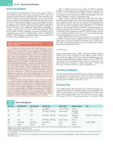
TABLE Table of Investigations
138.2
VWD Type vWF : RCo IU/dL a vWF : Ag IU/dL a RCo/Ag IU/dL a FVIII : C IU/dL a Multimer Pattern b Other
1 Low Low Equivalent ~1.5× VWF : Ag Normal
2A Low Low VWF : RCo < VWF : Ag Low or normal Abnormal
↓ HMWM
c
2B Low Low VWF : RCo < VWF : Ag Low or normal Abnormal ↑ RIPA (↓ platelet count)
↓ HMWM
2M Low Low VWF : RCo < VWF : Ag Low or normal Normal
2N Normal/low Normal/low Equivalent <30 Normal ↓ VWF : FVIIIB d
3 Absent Absent NA <10 Absent
a Relative to the reference range (approximate values); VWF : RCo (50–200 IU/dL); VWF : Ag (50–200 IU/dL); FVIII : C (50–150 IU/dL).
b HMWM, High-molecular-weight multimers.
c Increased agglutination at low concentrations of ristocetin.
d The ability of VWF to bind and protect FVIII is reduced. VWF and FVIII levels can look exactly like those in males with mild haemophilia A or in symptomatic hemophilia
A carrier females.
Ag, Antigen, FVIII : C, FVIII level; NA, not applicable; RCo, ristocetin cofactor; RIPA, ristocetin-induced platelet aggregation; VWD, von Willebrand disease; VWF:FVIIIB,
FVIII-binding assay.