
Page 515 - Hematology_ Basic Principles and Practice ( PDFDrive )
P. 515
Chapter 32 Acquired Disorders of Red Cell, White Cell, and Platelet Production 435
reduction in the variability of the CTL repertoire can occur in older constitutively express perforin and FasL and can suppress neutrophil
adults, and clonal or oligoclonal expansion of CTL populations may development in vitro. Typical clonal LGL cells seem to be terminally
be more frequent in older individuals. If asymptomatic, this disorder differentiated and cannot be effectively expanded in vitro by poly-
has been termed monoclonal clonopathy of unclear significance. clonal mitogens.
It is likely that a polyclonal CTL response predates the outgrowth
of the immunodominant T-LGL leukemia clone (see Fig. 32.3). The
PATHOGENESIS putative transforming event most likely involves a memory cell that
feeds into the mature effector CTL compartment. Under normal
Inciting Events physiologic circumstances, activated effector T cells are deleted after
antigen-driven expansion by Fas-mediated apoptosis. The failure of
T-LGL leukemia frequently arises in the context of a reactive poly- an activated memory and/or effector clone to undergo apoptosis may
clonal CTL expansion undergoing transformation in a manner similar result in its persistent expansion. LGL leukemia cells express high
to that proposed for CLL. It is possible also that in T-LGL leukemia levels of Fas/FasL, yet themselves are resistant to Fas-mediated apop-
one of the effector CTL clones may be initially driven by an inciting tosis. It is conceivable that persistent LGL leukemia cell expansion
antigen, may transform, and consequently the cells fail to undergo may result from this resistance to homeostatic apoptosis. In addition
apoptosis. The initial or initiating polyclonal response may be a to the high surface expression of Fas/FasL, soluble FasL has been
component of the pathophysiologic process associated with infectious detected in sera from T-LGL leukemia patients and may contribute
agents, rheumatoid arthritis, or other autoimmune disorders. to the induction of apoptosis of neutrophil precursors in the bone
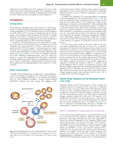
An initial T cell–mediated process may be responsible for cytope- marrow.
nias in the absence of clonal predominance. In concurrence with this An LGL clone persists mostly in the G 0/G 1 phase of cell cycle,
hypothesis, the clinical spectrum of T-LGL is determined by the and clonal transformation may also be because of a constitutive
specificity of the TCR: for example, if myeloid precursors are targets overexpression of prosurvival and antiapoptotic transcription factors.
of clonal CTL, neutropenia will be a clinical manifestation. Con- STAT3 has been shown to be involved in cellular transformation
versely, if erythroid progenitors are affected, patients will present with along with an active Src family kinase and appears to be constitutively
anemia (see Fig. 32.1 and Fig. 32.6). However, unlike the cytopenias activated in T-LGL leukemia cells. In addition, a constitutive activa-
that resolve following immunosuppression, the CTL clone may tion of an Src family kinase in T-LGL leukemia (likely Lck or Fyn)
persist at a certain level, suggesting that other disease mechanisms has been reported that may be related to this increased STAT phos-
220
involving soluble factors play a role in the development of the cyto- phorylation. It has been proposed that STAT3 activation in T-LGL
penias. Various soluble agents, including (FasL) and perforin, have may inhibit apoptosis downstream of Fas receptor signaling by
been implicated in the pathophysiology of the cytopenias in T-LGL induction of myeloid cell leukemia-1 (MCL1), a member of the
leukemia. B-cell lymphoma (BCL)2 family of antiapoptotic proteins. This
finding is further supported by data showing that blockade of STAT
signaling in T-LGL cells leads to the reversal of Fas resistance. Simi-
Clonal Transformation larly, constitutive activation of the extracellular signal-related kinase
mitogen-activated protein kinase pathway seems to play a role in
Clinically, T-LGL leukemia does not behave like a typical leukemia: survival of NK- and T-LGL leukemia cells.
excessive accumulation of malignant cells is often absent, and progres-
sion to a more malignant phenotype is rare. Instead, the expanded
clone in T-LGL leukemia resembles a normal antigen-activated Extreme Clonal Expansion and the Nonrandom Nature
+
+
CD8 CD57 effector cell; both normal and malignant LGL of the T-LGL
Molecular analysis of the TCR repertoire in T-LGL leukemia has
revealed a spectrum of expansion of the T-cell clone in individual
+
Clonal expansion patients. In some cases, up to 98% of the CD8 repertoire consists
of only one clone, a surprising finding given the absence of immu-
nodeficiency among T-LGL leukemia patients. In healthy controls,
even the most predominant clones, most likely reactive to ubiquitous
antigens, represent around 1% of the entire TCR repertoire. It is
possible that structurally similar clonotypes present in some patients
with T-LGL arise in the context of initial polyclonal CTL response
Cytokine inhibition and the initial transformation step is not random (see Fig. 32.3).
FasL Once a pathogenic immunodominant clonotype is identified and
IFN, TNF characterized, its sequence may be used for molecular tracking.
Erythroid TCR Genetic Alterations in Large Granular Lymphocyte
progenitor Direct cytotoxicity
No recurrent chromosomal aberrations or mutations have been found
Myeloid to be associated with LGL leukemia. Massively parallel second-
progenitor generation sequencing technology has been used successfully to
PRCA
uncover the genetic background of LGL leukemia. Whole exome
Neutropenia sequencing has aided in the discovery of somatic STAT3 mutation,
an oncogene located in chromosome 17 in 40% of the LGL cases.
The STAT3 missense mutations (D661V, D661Y, D661H, Y640F,
N647I, and K658N), as well as the insertion mutation (Y657_
K658insY), were located in the SH2 domain on the dimerization
Fig. 32.6 PATHOPHYSIOLOGY OF CYTOPENIAS IN T-CELL LARGE interface that mediates STAT3 activation. STAT3 mutations were
GRANULAR LYMPHOCYTE LEUKEMIA. FasL, Fas ligand; IFN, inter- detected in one-third cases of NK–LGL unifying T and NK cell
feron; PRCA, pure red cell aplasia; TCR, T-cell receptor; TNF, tumor necrosis lymphoproliferative pathogenesis. Several studies have identified
factor. STAT3 and STAT5b mutations specific to LGL at various frequencies.