
Page 516 - Hematology_ Basic Principles and Practice ( PDFDrive )
P. 516
436 Part IV Disorders of Hematopoietic Cell Development
Of interest, patients with STAT3 mutations were more likely to have may provide a rational target for immunosuppressive therapy. In
autoimmune conditions and neutropenia and tend to respond to MDS, T-LGL leukemia has been reported to negatively affect the
treatment. Sequencing of STAT3 wildtype cases revealed activating outcome of therapy directed against the CTL clone. LGL leukemia
mutations in STAT5b in 2% of the cases. STAT3 and STAT5b muta- has also been described in conjunction with hemolytic anemia fol-
tions can be used as molecular markers for LGL leukemia diagnostics, lowing bone marrow transplantation, perhaps a process initially
and they present novel therapeutic targets for STAT3 and STAT5b driven by an alloantigen or infectious agent such as EBV. As with
inhibitors. Interestingly not only STAT mutations were identified in infections, distinction between an LGL leukemia and reactive lym-
aplastic anemia and MDS cases with concomitant LGL but they were phoproliferation may be blurred. For example, neutropenia may be
small clones in 7% of cases without clinical evidence of LGL. associated with various degrees of clonality, with an LGL leukemia
representing the most extreme form of this process.
CLINICAL PRESENTATION AND PHYSICAL FEATURES
LABORATORY DIAGNOSIS
Patients with T-LGL leukemia present at a median age of about 55
years, with an equal male/female distribution. The clinical course may Diagnostic criteria remain a subject of considerable discussion (Table
be indolent and chronic. Patients are asymptomatic in one-third of 32.7). Traditionally LGL lymphocytosis (identified by morphologic
cases. The most common clinical presentation is neutropenia characteristics and flow cytometry) is a significant diagnostic crite-
(observed in approximately 85% of patients) often accompanied by rion. However, not all clonal cells display the typical morphologic
infections or neutropenic fever. However, in contrast to neutropenia features, and some patients present with leukopenia. Consequently
associated with other hematologic disorders, LGL leukemia patients an LGL count of greater than 2000/µL of blood has been abandoned
may remain surprisingly free of infectious complications for extended as a strict diagnostic requirement, and lower numbers such as
periods of time regardless of the depressed ANC. Despite the extreme 0.400/µL of blood have been proposed. 212,221 Most investigators
+
clonality within the T-cell population (suggesting a decreased antigen consider the presence of an expanded homogeneous CD3 , TCR-
+
+
−
+
+
recognition spectrum), opportunistic infections are rare. Other αβ , CD8 , CD16 , CD28 , CD57 cell population as diagnostic of
−
−
−
+
+
single-lineage cytopenias, including PRCA and immune-mediated T-LGL leukemia and CD2 , sCD3 , CD3ε , TCR-αβ , CD4 ,
+
+
+
thrombocytopenia, accompany T-LGL leukemia less frequently than CD8 , CD16 , CD56 , CD57 (variable) for NK-LGL leukemia. In
+
neutropenia. Pancytopenia may be related to splenomegaly reported almost all patients the expanded clone is CD8 (only very rarely
+
in 20% to 50% of patients. Hepatomegaly is present in a minority CD4 ), and in the majority of cases this population also expresses
of patients (10–20%). Lymphadenopathy and B symptoms may also CD57, but LGL leukemia cases without this marker have been
occur; however, this is uncommon. It has been reported that preg- observed. Clinical correlations based on immunophenotypic charac-
+
nancy can improve neutropenia in women with LGL leukemia. teristics have been defined; CD8 +(dim )/CD57 LGLs are associated
Clinical transformation to a more malignant form is rare. The clinical with clonal T-LGL leukemia and neutropenia, CD16 expression with
presentation of NK-cell LGL lymphocytosis is very similar to that complete or partial loss of CD5 is associated with T-LGL leukemia
+
seen in T-LGL leukemia with regard to lymphocyte counts, associated but not cytopenias, and CD8 +(dim )/CD57 with loss of CD5 expres-
conditions, treatment responses, and survival. sion is associated with T-LGL leukemia with severe neutropenia. In
addition, clinically aggressive T-LGL leukemia is characterized by
expression of CD26. In most cases of LGL leukemia, CD94 is
Clinical Overlap and Associations expressed at increased levels, and other receptors for class I MHC
molecules are abnormally expressed. Some investigators have sug-
+
In some clinical circumstances, natural or pathologic immune gested that a pool of CD8 memory cells exists that lack CD57
responses can resemble T-LGL expansions. For example, responses to expression but feed into the mature CD57 effector compartment.
viruses such as CMV or EBV, although of an oligoclonal or polyclonal The size of the abnormal clone defining T-LGL leukemia remains
nature, may display a strong clonal dominance mimicking at times a controversial. It is likely that the size of the leukemic T-cell popula-
true clonal process. Consequently, polarized CTL responses in the tion influences the detection of the clonal TCR γ-chain (G) rear-
context of infections have to be distinguished from true LGL leuke- rangement by PCR or Southern blotting; thus, such tests are
mia. T-LGL leukemia can occur concomitantly with several auto- considered mandatory for diagnosis. These methods may detect a
immune diseases. Rheumatoid arthritis is likely the most common clonal population that represents 15% of the cell population, but it
association, occurring in one-third of patients with T-LGL leukemia, is also likely that smaller CTL numbers may be consistent with latent
but additional diseases include ulcerative colitis, Sjögren syndrome, T-LGL detected only if more precise methods are used. T-LGL can
systemic lupus erythematosus, multiple sclerosis, and a number of
other (auto)immune conditions have been described. Felty syndrome
is characterized by neutropenia with rheumatoid arthritis and sple-
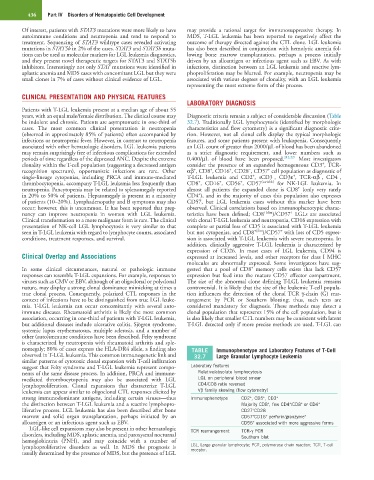
nomegaly; 80% of cases express the HLA-DR4 allele, a finding also TABLE Immunophenotype and Laboratory Features of T-Cell
observed in T-LGL leukemia. This common immunogenetic link and 32.7 Large Granular Lymphocyte Leukemia
similar patterns of cytotoxic clonal expansion with T-cell infiltration
suggest that Felty syndrome and T-LGL leukemia represent compo- Laboratory features
nents of the same disease process. In addition, PRCA and immune- Relative/absolute lymphocytosis
mediated thrombocytopenia may also be associated with LGL LGL on peripheral blood smear
lymphoproliferation. Clonal expansions that characterize T-LGL CD4/CD8 ratio reversed
leukemia can appear similar to oligoclonal CTL responses elicited by Vβ family skewing (flow cytometry)
strong immunodominant antigens, including certain viruses—thus Immunophenotype CD2 , CD5 , CD3 +
+
+
the distinction between T-LGL leukemia and a reactive lymphopro- Majority CD8 , few CD4 /CD8 or CD4 +
+
+
+
liferative process. LGL leukemia has also been described after bone CD27 CD28 −
+
marrow and solid organ transplantation, perhaps initiated by an CD57 CD16 perforin/granzyme +
+
+
alloantigen or an infectious agent such as EBV. CD56 associated with more aggressive forms
+
LGL-like cell expansions may also be present in other hematologic TCR rearrangement TCR-γ PCR
disorders, including MDS, aplastic anemia, and paroxysmal nocturnal Southern blot
hemoglobinuria (PNH), and may coincide with a number of
lymphoproliferative disorders as well. In MDS the prognosis is LGL, Large granular lymphocyte; PCR, polymerase chain reaction; TCR, T-cell
receptor.
usually determined by the presence of MDS, but the presence of LGL