
Page 811 - Hematology_ Basic Principles and Practice ( PDFDrive )
P. 811
Chapter 50 Disorders of Phagocyte Function 697
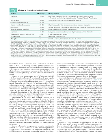
TABLE Infections in Chronic Granulomatous Disease
50.3
Infections Infections (%) Infecting Organisms
Pneumonia 70−80 Aspergillus, Staphylococcus, Burkholderia cepacia, Pseudomonas, Nocardia,
Mycobacterium (including atypical), Serratia, Candida, Klebsiella, Paecilomyces
Lymphadenitis 50−60 Staphylococcus, Serratia, Candida, Klebsiella, Nocardia
Cutaneous infections/impetigo 50−60
Hepatic or perihepatic abscesses 20−30 Staphylococcus, Serratia, Streptococcus viridans, Nocardia, Aspergillus
Osteomyelitis 20−30 Serratia, Aspergillus, Paecilomyces, Staphylococcus, B. cepacia, Pseudomonas, Nocardia
Perirectal abscesses or fistulae 15−30 Enteric gram-negative organisms, Staphylococcus
Septicemia 10−20 B. cepacia, Pseudomonas, Salmonella, Staphylococcus, Serratia, Klebsiella
Urinary tract infections or pyelonephritis 5−15 Enteric gram-negative organisms
Brain abscesses <5 Aspergillus, Staphylococcus
Meningitis <5 Candida lusitaniae, Haemophilus influenzae, B. cepacia
The relative frequencies of different types of infections in chronic granulomatous disease are estimated from data pooled from several large series of patients in the
United States, Europe, and Japan: (1) Mouy R, Fischer A, Vilmer E, et al: Incidence, severity, and prevention of infections in chronic granulomatous disease. J Pediatr
114:555, 1989; (2) Bemiller LS, Roberts DH, Starko KM, et al: Safety and effectiveness of long-term interferon gamma therapy in patients with chronic granulomatous
disease. Blood Cells Mol Dis 21:239, 1995; (3) Forrest CB, Forehand JR, Axtell RA, et al: Clinical features and current management of chronic granulomatous disease.
Hematol Oncol Clin North Am 2:253, 1988; (4) Hitzig WH, Seger RA: Chronic granulomatous disease, a heterogeneous syndrome. Hum Genet 64:207, 1983; (5) Tauber
AI, Borregaard N, Simons E, et al: Chronic granulomatous disease: A syndrome of phagocyte oxidase deficiencies. Medicine (Baltimore) 62:286, 1983; (6) Cohen MS,
Isturiz RE, Malech HL, et al: Fungal infection in chronic granulomatous disease. The importance of the phagocyte in defense against fungi. Am J Med 71:59, 1981;
(7) Hayakawa H, Kobayashi N, Yata J: Chronic granulomatous disease in Japan: A summary of the clinical features of 84 registered patients. Acta Paediatr Jpn 27:501,
1985; and (8) Johnston RB, Newman SL. Chronic granulomatous disease. Pediatr Clin North Am 24:365, 1977. These series encompass approximately 550 patients
with CGD after accounting for overlap between reports. Unpublished data from the United States CGD Registry encompassing 368 patients was also used to estimate the
relative frequencies of infections and the responsible organisms. The infecting organisms are arranged in approximate order of frequency for each type of infection. Note:
B. cepacia was previously classified as Pseudomonas cepacia.
brackish fresh water and which can cause a febrile illness with bacte- and the patient’s leukocytes. These lesions become granulomas as the
remia in CGD. A previously unknown gram-negative bacteria, host uses lymphocytes and activated macrophages to assist in contain-
Granulobacter bethesdensis, was recently identified in a CGD patient ing the pathogens. However, this complication is not always clearly
with recurrent fevers associated with chronic necrotizing deep lym- linked to persistent infection and in these cases, is speculated to
phatic infection. This organism is a member of the Acetobacteraceae involve a dysregulated inflammatory response, inefficient degradation
family, which has previously not been linked to invasive human of debris, or both. In the absence of oxidant production, excessive
disease. production of cytokines and delayed neutrophil apoptosis at inflam-
Pneumonia is the most common type of infection seen in CGD matory sites appear to contribute as underlying mechanisms.
with S. aureus, Aspergillus spp., B. cepacia, and enteric gram-negative As a result of persistent inflammatory stimulation, CGD patients
organisms as the major pathogens. It is noteworthy that B. cepacia can have a variety of more chronic complications (Table 50.4).
has emerged as a particularly lethal organism in CGD. This organism Lymphadenopathy, hepatosplenomegaly, eczematoid dermatitis, and
often is not covered with the first line of antibiotics used for S. aureus anemia of chronic disease (hemoglobin levels usually 8–10 g/dL) are
and most gram-negative organisms and can quietly proliferate (with common manifestations of this process and are most prominent in
persistent fevers) to the point of quick, explosive collapse caused by the first 5 to 10 years of life in those with CGD. Throughout the
endotoxic shock. Intravenous trimethoprim-sulfamethoxazole (TMP- body, granuloma formation can lead to dysfunction and obstruction
SMX) has been most effective in treating patients if given before in the esophagus, urinary bladder, and kidneys. In the stomach, the
widespread dissemination of the infection. Proven or suspected gastric antral narrowing can be severe enough in infants and children
Aspergillus infections were treated with amphotericin B therapy, but to resemble pyloric stenosis. Inflammatory involvement of the gastro-
new azole antifungal agents are now typically used. intestinal tract can be seen in up to one-third of CGD patients, typi-
Lymphadenitis is the second most common infection and is cally in association with the X-linked form. A chronic ileocolitis
usually caused by gram-negative organisms, S. aureus, or Serratia resembling Crohn disease occurs in about 10% of patients and can
marcescens. Incision and drainage should not be delayed if the lesion range from mild diarrhea to a debilitating syndrome of bloody diar-
fails to respond to parenteral antibiotics. Cutaneous abscesses should rhea and malabsorption that can necessitate a colectomy. Interestingly,
be similarly managed. Recurrent perinatal impetigo is almost a sig- antigliadin antibodies suggesting Crohn disease are positive in more
nature infection in CGD and often requires months of therapy than 50% of CGD patients. Other types of chronic inflammation
(mostly oral antibiotics) to clear. Hepatic (and perihepatic) abscesses include gingivitis, chorioretinitis, destructive white matter lesions in
are also common in CGD and are usually, but not always, caused by the brain, and glomerulonephritis. Discoid lupus has been reported
S. aureus. Most lesions require drainage (needle or surgical) to permit in 10% to 20% of patients, and occasional patients may develop
efficient healing to occur. Bone infections, most commonly caused systemic lupus erythematosus, sarcoidosis, or rheumatoid arthritis.
by Serratia spp. or Aspergillus spp., are particularly problematic in The underlying mechanisms are poorly defined, although recent
CGD and arise from either hematogenous or contiguous spread (as studies suggest that these manifestations may be partly related to
often is the case with Aspergillus infections in the lung invading the subtle defects in the absence of NADPH oxidase in memory B or
ribs, vertebral bodies, or the diaphragm). Perirectal abscesses are T cells.
difficult to treat, even with months of therapy, and can lead to fistula Carriers of CGD, whether the X-linked form or any one of the
formations. AR forms, are usually asymptomatic with two important exceptions.
Chronic inflammation with granuloma formation is a distinctive First, about one-fourth of X-linked carriers are at risk of developing
hallmark of CGD and contributes to some of its more problematic mild to moderately severe discoid lupus erythematosus characterized
complications. 8,14 In some cases, this results from imperfectly con- by discoid skin lesions and photosensitivity. 3,14 The onset is usually
trolled infections in which stalemates develop between the pathogen in the second decade of life. The disease does not progress to systemic