
Page 951 - Hematology_ Basic Principles and Practice ( PDFDrive )
P. 951
834 Part VII Hematologic Malignancies
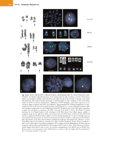
t(11;14)
A
t(8;14)
B
t(8;22)
C
t(14;18)
D
E F
Fig. 56.55 MOST FREQUENT CHROMOSOMAL ABNORMALITIES IN NON-HODGKIN LYM-
PHOMA. (A) Partial G-banded karyotype of t(11;14)(q13;q32.3) (left), resulting in CCND1-IGH fusion
(yellow) on metaphase (middle) and in interphase cell (right) present in the majority of patients with mantle
cell lymphoma. (B) Partial G-banded karyotype of t(8;14)(q24;q32.3) (left), resulting in MYC-IGH fusion
[yellow on der(8) on isolated chromosomes]. Application of MYC breakapart probe shows separation of red
and green signals consistent with MYC rearrangement. Approximately 80% of Burkitt lymphomas are char-
acterized by t(8;14). (C) Partial G-banded karyotype of t(8;22)(q24;q11), a variant of Burkitt lymphoma (left).
Two interphase lymph node cells after hybridization with MYC breakapart probe and CEP8 (aqua). Separation
of green and red signals is consistent with MYC relocation from 8q24 to 22q11. (D) Partial G-banded
karyotype of t(14;18)(q32.3;q21.3) (left), resulting in IGH-BCL fusion (two yellow signals, middle). A
composite image on the right shows both a partial karyotype and FISH study of triplicated der(18)t(14;18)
and three copies of IGH-BCL2 fusion (yellow), as well as normal chromosome 14 and 18. Multiple copies of
abnormal der(18) chromosome are associated with progressive disease similar to a duplication of the Philadel-
phia chromosome in the blast crisis of chronic myelogenous leukemia. (E) Lymph node cell from a patient
with diffuse large B-cell lymphoma, showing four copies of BCL6 (breakapart probe). BCL6 is localized at
3q27 and is numerically or structurally rearranged in 35% of diffuse large B-cell lymphoma. (F) Bone marrow
metaphase and interphase cell hybridized with breakapart MALT1 gene at 18q21 (left) indicating two fusion
(yellow) signals when MALT1 gene is intact. In contrast, rearrangement of the MALT1 gene in MALT lym-
phoma usually is the consequence of t(11;18)(q21;q21.1), as shown in the cell (right) with clear separation of
the 3′ end (green) and the 5′ end (red).